Short Answer
Definition of Magnetic Resonance Imaging (MRI)
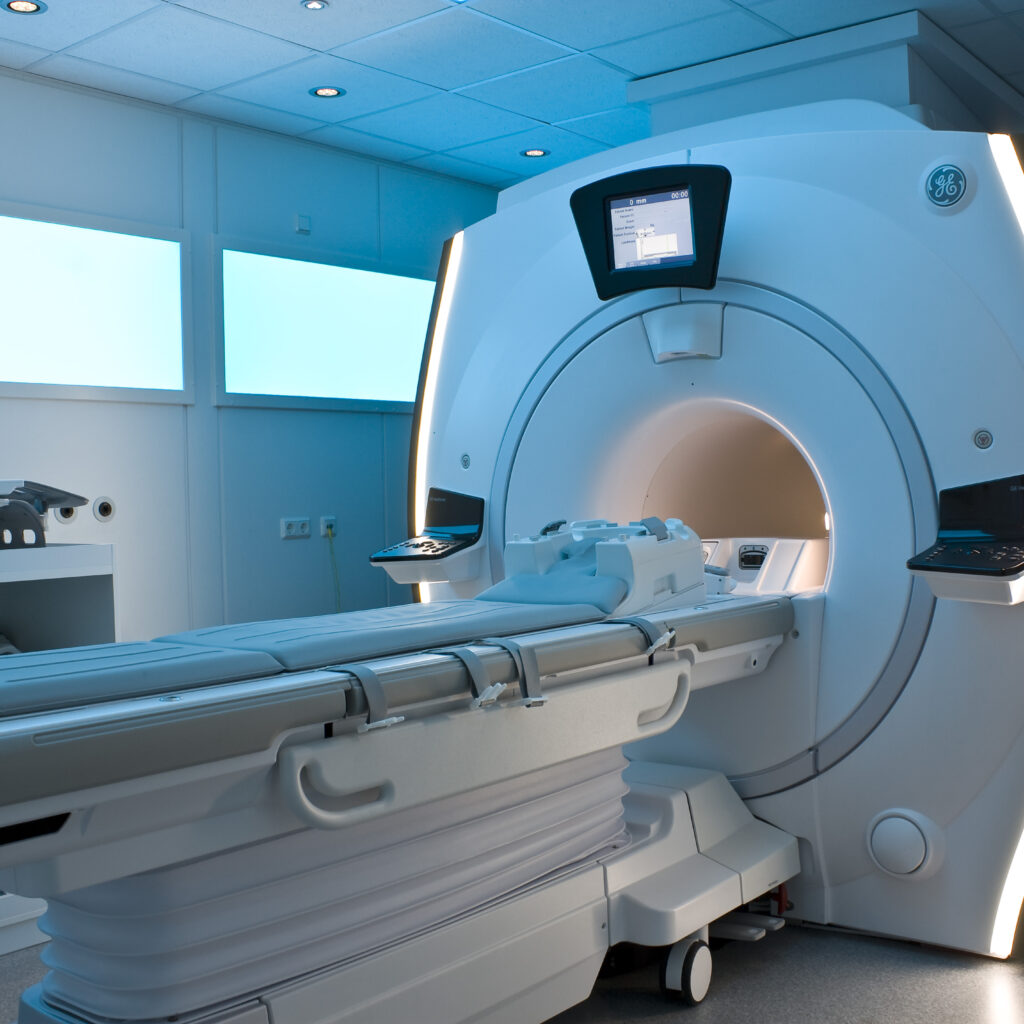
Magnetic Resonance Imaging (MRI) is a sophisticated medical imaging technique that utilizes the principles of nuclear magnetic resonance (NMR) to generate detailed images of the internal structures of the body. It is a non-invasive diagnostic tool that produces high-resolution images, particularly effective for soft tissues, without exposing patients to ionizing radiation.
Historical Background and Scientific Foundations
The conceptual roots of MRI trace back to the 1940s, a transformative era in quantum mechanics and atomic physics. Physicist Isidor Isaac Rabi was pivotal in this period, demonstrating that atomic nuclei behave like minuscule magnets that align and respond to external magnetic fields. His groundbreaking research earned him the Nobel Prize in Physics in 1944 and laid the essential groundwork for future imaging technologies.
During the 1950s, further advancements were made by Felix Bloch and Edward Purcell, who independently developed methods to measure the magnetic properties of atomic nuclei. Their work enabled scientists to investigate molecular and atomic structures non-destructively, initially benefiting chemical research. However, the application of these principles to medical imaging was not yet realized.
Development of MRI Technology
The transition from theoretical NMR to practical medical imaging occurred in the 1970s, largely due to the efforts of Raymond Damadian, an American physician and researcher. Damadian proposed that cancerous tissues exhibit distinct NMR signals compared to healthy tissues, a hypothesis that motivated him to build the first MRI scanner, nicknamed the “Indomitable.” In 1971, this device produced the earliest images of internal body anatomy, marking a significant milestone in medical diagnostics.
Subsequent technological enhancements were driven by innovators such as Dr. John Mallard and Dr. George K. S. Hounsfield, the latter known for inventing the computed tomography (CT) scan. By integrating advanced computational algorithms with NMR principles, these pioneers improved image clarity and reduced scanning durations, overcoming critical barriers to clinical adoption.
Principles and Mechanism of MRI
MRI operates by aligning the magnetic moments of hydrogen nuclei in the body using a strong external magnetic field. Radiofrequency pulses then perturb this alignment, and as the nuclei return to their equilibrium state, they emit signals that are detected and translated into images. The contrast in these images arises from variations in tissue properties such as proton density and relaxation times.
Formula and Mathematical Explanation
The fundamental physics of MRI can be described by the Larmor equation:
ω = γB
- ω (Larmor frequency): The precession frequency of the nuclear spins.
- γ (Gyromagnetic ratio): A constant specific to the type of nucleus (e.g., hydrogen).
- B (Magnetic field strength): The intensity of the external magnetic field applied.
This relationship governs the resonance condition necessary for nuclei to absorb and emit radiofrequency energy, which is fundamental to image formation.
Clinical Applications and Real-World Examples
Since its clinical introduction in the 1980s, MRI has become indispensable in diagnosing a wide range of conditions. It excels in imaging the brain, spinal cord, joints, and abdominal organs, providing unparalleled soft tissue contrast. MRI assists not only in visualizing anatomical structures but also in guiding treatment plans and surgical procedures.
The advent of functional MRI (fMRI) has expanded MRI’s utility by enabling the mapping of brain activity through blood flow changes, significantly advancing research in neurology, psychology, and cognitive sciences.
Safety Considerations and Ethical Aspects
Unlike imaging modalities that use ionizing radiation, such as X-rays and CT scans, MRI employs strong magnetic fields and radio waves, which do not carry the same risks of radiation exposure. This safety profile has made MRI a preferred choice for repeated imaging, especially in vulnerable populations. Nonetheless, the presence of strong magnetic fields necessitates careful screening for metallic implants and devices to prevent adverse effects.
Challenges and Technological Evolution
Despite its advantages, MRI technology initially faced hurdles including high costs, complex operation, and limited accessibility in smaller healthcare settings. These challenges spurred interdisciplinary collaborations among physicists, engineers, and medical professionals to refine MRI systems, improve affordability, and enhance user-friendliness.
Continuous innovation has led to diverse MRI modalities tailored to specific diagnostic needs, ensuring its role as a cornerstone of modern medical imaging.
Common Misconceptions About MRI
MRI uses harmful ionizing radiation.
MRI employs magnetic fields and radio waves, which do not involve ionizing radiation, making it safer than X-rays or CT scans.
MRI scans are painful or invasive.
MRI is a non-invasive and painless procedure, though some patients may experience discomfort due to confined spaces or noise.
Significance of MRI in Medicine and Science
The invention and evolution of MRI represent a remarkable fusion of physics, engineering, and medical science. This technology has revolutionized diagnostic imaging by providing detailed insights into the human body’s internal structures without invasive procedures or radiation risks. MRI’s ability to enhance diagnosis, treatment planning, and research underscores its vital role in advancing healthcare and deepening our understanding of human biology.
Conclusion
The story of MRI is a testament to human creativity, perseverance, and interdisciplinary collaboration. From its origins in quantum physics to its current status as a medical imaging cornerstone, MRI exemplifies how scientific inquiry can translate into transformative technologies that improve human health. As MRI continues to evolve, it remains a powerful symbol of the ongoing quest to illuminate the complexities of the human body and enhance the quality of life.
FAQ
What is MRI?
MRI stands for Magnetic Resonance Imaging, a non-invasive medical imaging technique that creates detailed images of the body's internal structures.
How does MRI work?
MRI works by aligning the magnetic moments of hydrogen nuclei in the body using a strong magnetic field and radiofrequency pulses, which generate signals that are converted into images.
Is MRI safe?
Yes, MRI is considered safe as it does not use ionizing radiation, but it requires careful screening for metallic implants.


Leave a Reply