Short Answer
Overview of Radiology and Medical Imaging Physics
Radiology and medical imaging represent essential branches within healthcare, enabling the visualization, diagnosis, and management of numerous medical conditions. The integration of physics principles into these disciplines provides a foundation for understanding how imaging technologies function, driving innovations that enhance patient care and diagnostic accuracy. This article delves into the fundamental physics behind various imaging modalities, highlighting their significance and applications.
Definition and Scope of Medical Imaging
Medical imaging refers to the techniques and processes used to create visual representations of the interior of a body for clinical analysis and medical intervention. It allows healthcare professionals to observe internal structures non-invasively, facilitating early detection and treatment of diseases.
- Non-invasive visualization:
Medical imaging enables the examination of internal organs and tissues without surgical procedures. - Diagnostic enhancement:
It improves the accuracy and speed of diagnosing various health conditions. - Monitoring and treatment planning:
Imaging supports ongoing assessment and guides therapeutic decisions.
Primary Medical Imaging Modalities and Their Physical Principles
Several imaging techniques dominate clinical practice, each relying on distinct physical phenomena. Understanding these principles is crucial for optimizing image quality and ensuring patient safety.
X-ray Radiography
X-ray radiography is a widely utilized imaging method characterized by its quick execution and ease of interpretation. It operates by emitting ionizing radiation that passes through the body, producing images based on the differential absorption of X-rays by various tissues. Dense structures like bones absorb more radiation, appearing lighter on the resulting image, whereas softer tissues absorb less and appear darker.
Key physics concepts involved include photon energy, absorption coefficients, and scattering effects. Mastery of these factors is essential to balance image clarity with minimizing radiation exposure. The advent of digital radiography has further refined image processing, enhancing diagnostic precision beyond traditional visual assessments.
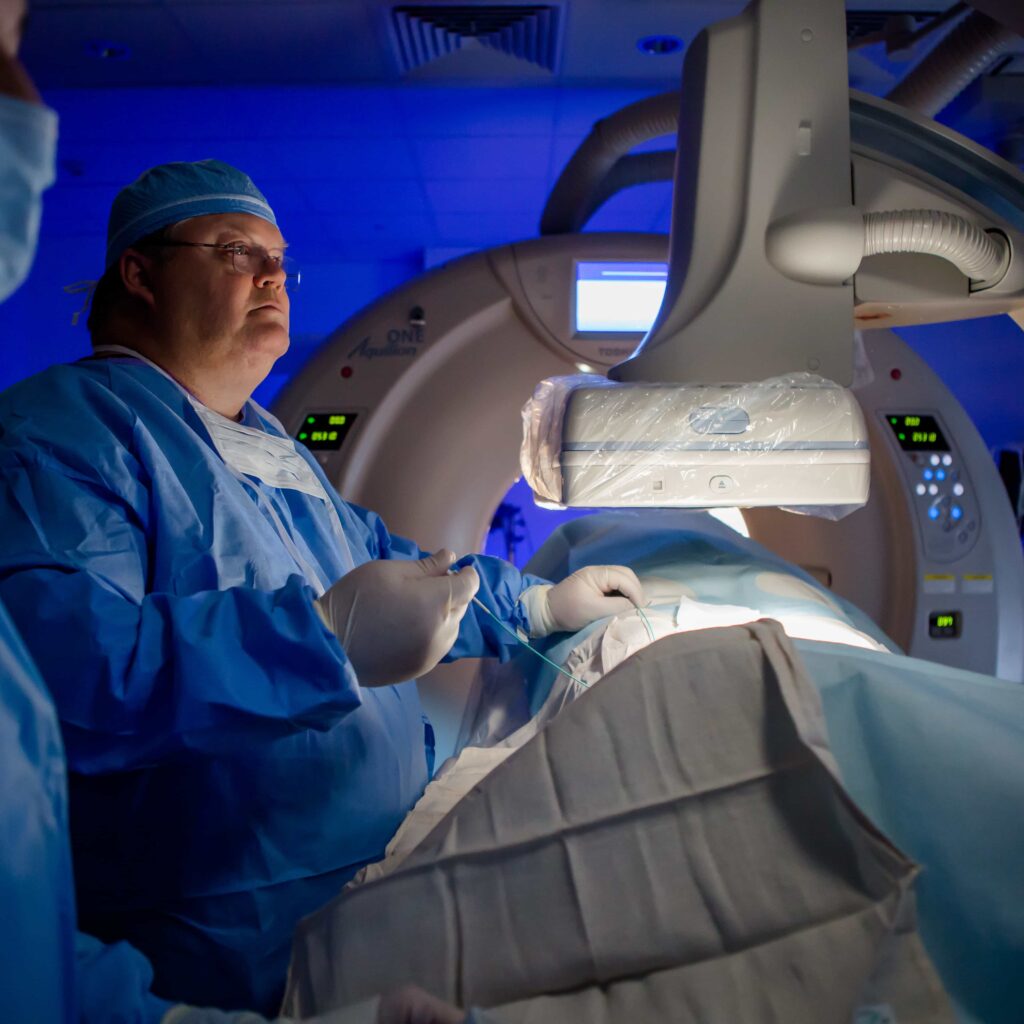
Computed Tomography (CT)
CT imaging builds upon X-ray technology by capturing multiple X-ray images from different angles around the patient. These images are then computationally reconstructed into detailed cross-sectional views using mathematical algorithms such as the Fourier transform. The technique relies heavily on understanding attenuation coefficients to differentiate tissue densities accurately.
Modern CT scanners, including multi-slice systems, provide rapid acquisition of high-resolution images, significantly improving diagnostic capabilities. However, the increased radiation dose associated with these advanced scans necessitates careful evaluation of risks versus benefits, prompting ongoing refinement of imaging protocols to prioritize patient safety.
Magnetic Resonance Imaging (MRI)
MRI distinguishes itself by utilizing strong magnetic fields and radiofrequency waves to generate images, rather than ionizing radiation. The underlying physics is based on nuclear magnetic resonance (NMR), where atomic nuclei in the body respond to magnetic fields, producing signals that are converted into detailed images.
This modality excels in soft tissue contrast, making it invaluable for detecting tumors, neurological disorders, and musculoskeletal abnormalities. Critical parameters such as relaxation times (T1 and T2) influence image quality and are manipulated to optimize visualization. MRI’s lack of ionizing radiation contributes to its favorable safety profile, making it a preferred diagnostic tool in many scenarios.
Ultrasound Imaging
Ultrasound employs high-frequency sound waves to create real-time images of soft tissues. It is extensively used in obstetrics, gynecology, and cardiology. The physics behind ultrasound involves wave propagation, reflection, refraction, and the Doppler effect, which allows assessment of blood flow dynamics.
Understanding acoustic impedance-the resistance sound waves encounter when passing through different tissues-is vital for image optimization. The portability and affordability of ultrasound devices enhance their clinical utility. Recent technological advances have introduced 3D and 4D imaging capabilities, enabling dynamic visualization of anatomical structures and fetal movements.
Nuclear Medicine
Nuclear medicine focuses on the use of radioactive substances (radiopharmaceuticals) to evaluate physiological functions within the body. Techniques such as positron emission tomography (PET) and single-photon emission computed tomography (SPECT) detect gamma rays emitted during radioactive decay, providing insights into metabolic activity and disease progression.
Fundamental physics concepts include radioactive half-life, decay chains, and radiation safety protocols. The fusion of molecular biology with imaging has led to the development of targeted radiotracers, enhancing diagnostic specificity and enabling personalized treatment monitoring. This modality uniquely visualizes biochemical processes in vivo, advancing precision medicine.
Physics Behind Medical Imaging Technologies
The effectiveness of medical imaging hinges on the application of various physical laws and phenomena:
- Ionizing Radiation:
Used in X-rays and CT scans, it involves photons with enough energy to ionize atoms, enabling tissue differentiation based on absorption. - Magnetic Resonance:
Relies on nuclear spin properties and magnetic fields to generate signals from atomic nuclei. - Acoustic Waves:
Ultrasound uses sound wave reflection and transmission to form images. - Radioactive Decay:
Nuclear medicine exploits the emission of radiation from unstable isotopes to assess physiological functions.
Mathematical Foundations in Imaging
Several mathematical tools underpin image reconstruction and analysis:
- Fourier Transform:
Converts spatial data into frequency components, essential in CT image reconstruction. - Attenuation Coefficient (μ):
Represents the rate at which X-rays are absorbed or scattered by tissues, influencing image contrast. - Relaxation Times (T1 and T2):
Describe the time constants for nuclear spin relaxation in MRI, affecting image contrast and quality.
Practical Applications and Clinical Impact
Medical imaging technologies have transformed healthcare by enabling:
- Early Disease Detection:
Identifying conditions such as cancer, cardiovascular diseases, and neurological disorders at treatable stages. - Guided Interventions:
Assisting in surgical planning and minimally invasive procedures. - Therapeutic Monitoring:
Tracking treatment efficacy and disease progression over time. - Personalized Medicine:
Tailoring treatments based on individual physiological and molecular imaging data.
Common Misunderstandings in Medical Imaging Physics
- Misconception: MRI uses harmful ionizing radiation.
Correction: MRI employs magnetic fields and radio waves, which do not involve ionizing radiation, making it safer for patients. - Misconception: Higher radiation doses in CT scans always yield better images.
Correction: While increased dose can improve image quality, it also raises patient risk; optimal protocols balance image clarity with safety. - Misconception: Ultrasound can image all body parts equally well.
Correction: Ultrasound is limited by bone and air interference, making it less effective for imaging certain organs like the lungs or brain.
Significance of Radiology and Medical Imaging Physics
The fusion of physics with medical imaging has revolutionized modern medicine, offering unparalleled insights into human anatomy and physiology. These technologies not only enhance diagnostic accuracy but also reduce the need for invasive procedures, improving patient comfort and outcomes. As research progresses, the synergy between physics and biology promises continual advancements, expanding the horizons of personalized healthcare and therapeutic innovation.
FAQ
What are the advantages of MRI over CT scans?
MRI provides better soft tissue contrast without using ionizing radiation, making it safer for repeated imaging and particularly useful for brain, muscle, and tumor imaging.
How does ultrasound imaging work?
Ultrasound uses high-frequency sound waves that reflect off tissues to create dynamic images, useful for soft tissue visualization and blood flow assessment.
Why is radiation dose a concern in some imaging modalities?
Because ionizing radiation can damage tissues and increase cancer risk, minimizing dose while maintaining image quality is critical in X-ray, CT, and nuclear medicine.
What advancements have improved CT imaging?
Technologies like multi-slice scanners and advanced reconstruction algorithms have enhanced CT image resolution and acquisition speed.


Leave a Reply