Short Answer
Definition of Functional Magnetic Resonance Imaging (fMRI)
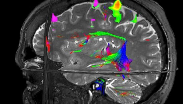
Functional Magnetic Resonance Imaging (fMRI) is an advanced neuroimaging technique that enables the visualization of brain activity by detecting changes in blood flow. Unlike conventional imaging methods that use ionizing radiation, fMRI employs strong magnetic fields and radiofrequency waves to non-invasively monitor neural function. This technology has become a cornerstone in both neuroscience research and clinical diagnostics, offering detailed insights into the brain’s functional architecture.
Principles Behind fMRI Technology
The operation of fMRI is grounded in the hemodynamic response, a physiological process where increased neuronal activity leads to elevated oxygen consumption and subsequent changes in local blood flow. When neurons in a specific brain region become active, they demand more oxygen, which is delivered via the bloodstream. fMRI detects these variations by measuring the Blood Oxygen Level Dependent (BOLD) signal, which reflects the ratio of oxygenated to deoxygenated hemoglobin. This mechanism allows for real-time mapping of brain function during cognitive or motor tasks without the need for invasive procedures.
Applications and Significance in Neuroscience and Medicine
fMRI has profoundly impacted our understanding of the brain’s complex functions. It facilitates the exploration of cognitive processes such as decision-making, emotional regulation, and behavioral responses. Clinically, fMRI is invaluable for preoperative planning, helping neurosurgeons identify and preserve critical brain regions during tumor removal or epilepsy surgery. Additionally, it plays a pivotal role in psychiatric research by uncovering neural patterns associated with disorders like depression, anxiety, and schizophrenia, thereby guiding personalized treatment strategies.
Safety Considerations and Potential Health Risks
While fMRI is widely regarded as a safe imaging modality, several safety aspects warrant attention:
- Magnetic Field Interactions:
The powerful static magnetic fields used in fMRI can pose risks for patients with ferromagnetic implants, pacemakers, or certain neurostimulators, as these devices may malfunction or move due to magnetic forces. - Claustrophobia and Anxiety:
The enclosed environment of the MRI scanner can induce psychological distress in some individuals, potentially triggering stress responses that affect overall well-being during the procedure. - Radiofrequency (RF) Exposure:
RF pulses are employed to generate images, and although current evidence suggests minimal biological impact, the long-term effects of repeated or prolonged exposure remain under investigation.
Technological Innovations to Enhance Patient Comfort
To address challenges such as claustrophobia, advancements like open MRI systems have been developed. These designs offer a more spacious scanning environment, reducing patient anxiety and improving the overall experience without compromising image quality. Such innovations are crucial for expanding the accessibility and tolerability of fMRI examinations.
Role of fMRI in Neurodegenerative Disease Diagnosis and Monitoring
As the global population ages, neurodegenerative conditions such as Alzheimer’s and Parkinson’s disease are becoming increasingly prevalent. fMRI contributes to early diagnosis and monitoring by detecting functional changes in brain regions affected by these disorders. However, the necessity for repeated imaging sessions raises questions about cumulative exposure and its implications, emphasizing the need for ongoing research into long-term safety.
Neuroplasticity and Rehabilitation Insights from fMRI
One of the most promising aspects of fMRI lies in its ability to reveal neuroplasticity-the brain’s capacity to reorganize and form new neural connections. This insight is particularly valuable in rehabilitation following strokes or traumatic brain injuries, where fMRI guides targeted therapies by identifying compensatory brain regions. Such applications underscore the technology’s therapeutic potential beyond diagnostics.
Ethical Considerations in the Use of fMRI
The expanding use of fMRI raises important ethical questions, especially regarding privacy and data security. As the technique can potentially decode neural correlates of thoughts and intentions, safeguarding sensitive brain data is paramount. Ethical frameworks must evolve to regulate the collection, sharing, and application of neuroimaging information, ensuring that fMRI serves to enhance human health without infringing on individual rights.
Common Misconceptions About fMRI
fMRI exposes patients to harmful ionizing radiation.
fMRI uses magnetic fields and radio waves, which do not involve ionizing radiation, making it safer than X-rays or CT scans.
The magnetic field in fMRI can cause implants to move inside the body.
While strong magnetic fields can affect ferromagnetic materials, patients with incompatible implants are screened out to prevent such risks.
fMRI can read thoughts directly.
fMRI measures brain activity patterns but cannot decode specific thoughts or intentions with certainty.
Why fMRI is Crucial in Modern Medicine and Research
Functional MRI stands as a transformative tool in both clinical and research domains. Its ability to non-invasively map brain activity has revolutionized diagnostics, treatment planning, and our fundamental understanding of neural processes. By enabling personalized medicine approaches and advancing neuropsychiatric research, fMRI contributes significantly to improving patient outcomes and expanding scientific knowledge. Continued innovation and ethical vigilance will ensure that its benefits are maximized while minimizing potential risks.
FAQ
What is fMRI?
fMRI is a neuroimaging method that detects brain activity by measuring changes in blood flow using magnetic fields and radio waves.
Are there any health risks associated with fMRI?
While fMRI is considered safe, risks include potential interference with implants, claustrophobia, and unknown long-term effects of repeated scans.
How does fMRI differ from other imaging techniques?
Unlike X-rays or CT scans, fMRI does not use ionizing radiation but instead relies on magnetic fields to visualize brain function.


Leave a Reply