Short Answer
Definition of Radiology Images
Radiology images refer to a broad spectrum of sophisticated imaging techniques used by healthcare professionals to visualize the internal anatomy and physiological functions of the human body. These images are generated through various technological methods, each designed to highlight different tissues, organs, or biological processes. The terminology for radiology images varies depending on the specific imaging modality employed, including terms such as “X-ray,” “computed tomography (CT) scan,” “magnetic resonance imaging (MRI),” “ultrasound,” and “nuclear medicine scans.”
- X-ray:
Uses electromagnetic radiation to capture images primarily of dense structures like bones. - CT scan:
Combines multiple X-ray images to create detailed cross-sectional views of the body. - MRI:
Utilizes magnetic fields and radio waves to produce high-resolution images of soft tissues. - Ultrasound:
Employs high-frequency sound waves to generate real-time images, especially useful in obstetrics and cardiology. - Nuclear Medicine Scans:
Involve radioactive tracers to assess organ function and detect abnormalities.
Fundamental Imaging Modalities and Their Mechanisms
X-ray Imaging
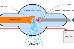
X-ray imaging is one of the most widely recognized radiological techniques. It operates by directing high-frequency electromagnetic waves through the body, which are absorbed differently by various tissues. Dense materials such as bones absorb more X-rays and appear white on the resulting image, while softer tissues allow more X-rays to pass through, appearing darker. This method is crucial in emergency medicine for quickly identifying fractures, dislocations, and foreign objects. Modern advancements include digital X-rays, which enhance image clarity and reduce radiation exposure.
Computed Tomography (CT) Scans
CT scans enhance traditional X-ray imaging by capturing multiple images from different angles around the body. These images are processed by computer algorithms to produce detailed cross-sectional “slices,” offering comprehensive views of soft tissues, organs, and blood vessels. This modality is invaluable for diagnosing complex conditions such as tumors, internal hemorrhages, and infections. The terminology “computed axial tomography (CAT) scan” is also used, reflecting its advanced computational approach. Additionally, 3D reconstructions from CT data provide even more detailed anatomical insights.
Magnetic Resonance Imaging (MRI)
MRI technology uses powerful magnetic fields combined with radiofrequency pulses to generate detailed images, particularly of soft tissues like the brain, muscles, and joints. Unlike X-rays and CT scans, MRI does not involve ionizing radiation, making it safer for repeated use. It is especially effective in diagnosing neurological disorders, musculoskeletal injuries, and soft tissue abnormalities. The images produced are often referred to as “MRI scans” or “MR images.”
Ultrasound Imaging
Ultrasound, or sonography, employs high-frequency sound waves to create live images of internal organs and blood flow. This technique is widely used in prenatal care, cardiac evaluations, and abdominal examinations. Its portability, real-time imaging capability, and absence of ionizing radiation make it a versatile and safe diagnostic tool. Ultrasound images are commonly called “ultrasonic images” or simply “ultrasounds.”
Nuclear Medicine Imaging
Nuclear medicine involves administering radiopharmaceuticals that emit gamma rays detectable by specialized cameras. This approach provides functional information about organs and tissues by tracking the distribution of radioactive tracers. Common nuclear imaging techniques include scintigraphy and positron emission tomography (PET). These scans are essential for assessing thyroid function, cardiac perfusion, and cancer detection. PET combined with CT (PET/CT) merges anatomical and functional data, enhancing diagnostic precision in oncology.
Hybrid Imaging Technologies
Hybrid imaging represents a cutting-edge advancement by integrating multiple imaging modalities to improve diagnostic accuracy. Examples include PET/CT and single-photon emission computed tomography combined with CT (SPECT/CT). These fusion imaging techniques combine the anatomical detail of CT with the functional insights of nuclear medicine scans, offering a comprehensive view of disease processes. Such integrated imaging is pivotal in personalized treatment planning and monitoring therapeutic responses.
Clinical Applications and Importance
Radiology images are indispensable in modern healthcare, providing critical information that guides diagnosis, treatment, and patient management. From emergency trauma assessment with X-rays to detailed neurological evaluations with MRI, each imaging modality contributes uniquely to medical decision-making. Nuclear medicine scans add a functional dimension, revealing physiological changes that anatomical images alone cannot detect. The continuous evolution of imaging technologies enhances the ability of clinicians to deliver precise, timely, and individualized care.
Common Misconceptions About Radiology Imaging
All radiology images expose patients to harmful radiation.
While X-rays, CT scans, and nuclear medicine involve ionizing radiation, MRI and ultrasound do not, making them safer alternatives in many cases.
Radiology images only show bones.
Many imaging modalities, such as MRI and ultrasound, provide detailed views of soft tissues, organs, and physiological functions.
Nuclear medicine scans are only used for cancer.
These scans are also vital for evaluating cardiac health, thyroid function, and other organ systems.
FAQ
What is the technical term for an X-ray image?
It is commonly called a radiograph or X-ray film.
How does a CT scan differ from a traditional X-ray?
A CT scan combines multiple X-ray images from different angles to produce cross-sectional slices of the body.
Why is MRI preferred for soft tissue imaging?
MRI uses magnetic fields and radio waves to create detailed images without ionizing radiation, making it ideal for soft tissues.
What is the role of nuclear medicine scans?
They provide functional information about organs by tracking radiopharmaceutical distribution within the body.
What are hybrid imaging techniques?
Hybrid imaging, such as PET/CT, merges anatomical and functional imaging to enhance diagnostic accuracy.


Leave a Reply