Short Answer
Definition of MRI Shimming
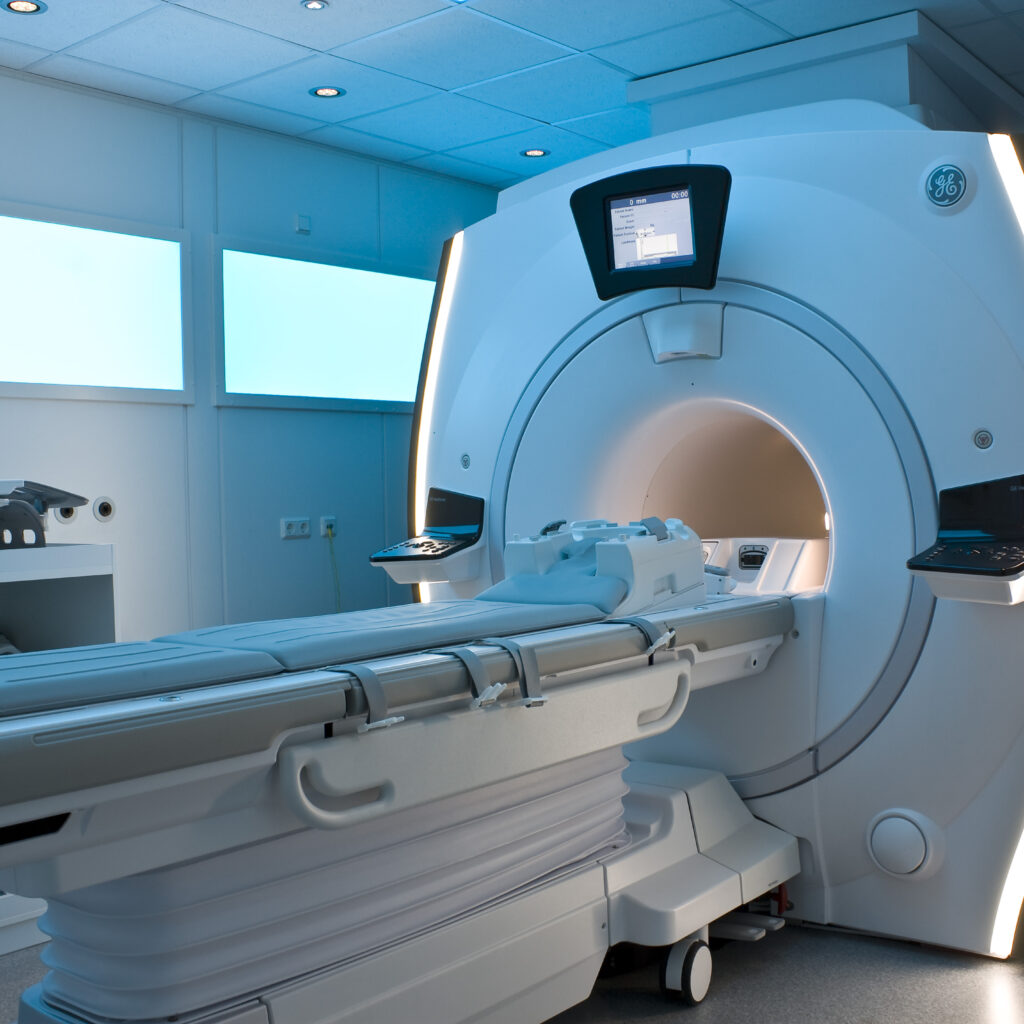
Magnetic Resonance Imaging (MRI) shimming is a critical process aimed at improving the uniformity of the magnetic field within an MRI scanner. This enhancement is essential for producing high-quality, accurate images of the human body without invasive procedures. Shimming corrects irregularities in the magnetic field, which can otherwise cause distortions and reduce image clarity.
Fundamentals of MRI Technology
MRI technology is based on the principles of nuclear magnetic resonance (NMR). It primarily detects signals from hydrogen nuclei found in water molecules throughout the body. When exposed to a strong magnetic field, these nuclei align and, upon excitation by radiofrequency pulses, emit signals that are captured and converted into detailed anatomical images. For optimal image resolution, the magnetic field must be highly uniform, maintaining consistent strength throughout the scanning area.
Sources of Magnetic Field Inhomogeneity
In practice, achieving a perfectly homogeneous magnetic field is challenging. Variations, known as field inhomogeneities, can arise from several factors:
- Scanner Design:
The physical components and construction of the MRI machine can introduce magnetic inconsistencies. - Patient Anatomy:
Differences in body composition and shape affect the magnetic environment inside the scanner. - External Magnetic Materials:
Nearby ferromagnetic objects or implants can distort the magnetic field.
These irregularities can lead to image artifacts, reduced contrast, and potential misdiagnoses.
Techniques of MRI Shimming
MRI shimming encompasses methods to correct magnetic field distortions, thereby enhancing image quality. The two primary approaches are passive and active shimming.
Passive Shimming
This technique involves placing ferromagnetic materials strategically within the MRI bore. These materials are selected and positioned to counterbalance the magnetic field irregularities by inducing opposing magnetic distortions. Although passive shimming can improve field uniformity, it is generally less flexible and less precise compared to active methods.
Active Shimming
Active shimming employs electromagnetic coils that dynamically adjust the magnetic field in real time. By modulating electrical currents through these coils, the system generates compensatory magnetic fields that neutralize inhomogeneities. This approach allows for fine-tuned, adaptable correction, resulting in superior magnetic field homogeneity.
Importance of MRI Shimming in Diagnostic Imaging
Effective shimming significantly enhances the diagnostic capabilities of MRI by:
- Improving Image Resolution:
Uniform magnetic fields produce clearer, more detailed images, enabling the detection of subtle abnormalities. - Reducing Scan Duration:
Better field homogeneity can shorten examination times, increasing patient comfort and minimizing motion artifacts. - Facilitating Accurate Diagnoses:
Enhanced image quality supports more reliable interpretation by radiologists and clinicians.
Challenges and Considerations in MRI Shimming
Despite its advantages, MRI shimming presents several challenges:
- Technical Complexity:
Calibrating shimming parameters requires specialized knowledge of electromagnetic principles and MRI physics. - Patient Variability:
Anatomical differences among patients can complicate shimming adjustments, necessitating individualized calibration for optimal results.
Future Directions in MRI Shimming
Ongoing advancements in MRI technology are driving innovations in shimming techniques. Emerging research focuses on integrating artificial intelligence and machine learning algorithms to automate and optimize active shimming processes. These developments promise to enhance precision, reduce operator dependency, and further improve diagnostic accuracy.
Summary
MRI shimming is an indispensable process that refines the magnetic field uniformity within MRI scanners, directly impacting image quality and diagnostic effectiveness. By addressing magnetic field inconsistencies through passive and active methods, shimming enables clearer visualization of anatomical structures and supports better clinical outcomes. As technology evolves, the refinement of shimming techniques will continue to play a pivotal role in advancing medical imaging and patient care.
FAQ
What is MRI shimming?
MRI shimming is the process of adjusting the magnetic field within an MRI scanner to make it more uniform, which improves image quality and diagnostic accuracy.
What are the main types of MRI shimming?
The two main types are passive shimming, which uses ferromagnetic materials to correct the field, and active shimming, which uses electromagnetic coils to dynamically adjust the magnetic field.
Why is MRI shimming important?
Shimming improves image resolution, reduces scan time, and facilitates accurate diagnoses by correcting magnetic field distortions.
What challenges exist in MRI shimming?
Challenges include technical complexity in calibration and variability in patient anatomy that requires individualized adjustments.
How is artificial intelligence used in MRI shimming?
AI and machine learning are being developed to automate and optimize active shimming processes, enhancing precision and reducing operator dependence.


Leave a Reply