Short Answer
Definition of Magnetic Resonance Imaging (MRI)
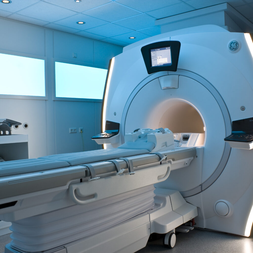
Magnetic Resonance Imaging (MRI) is an advanced diagnostic imaging technique that has transformed medical practice by providing detailed views of the body’s internal anatomy without exposing patients to ionizing radiation. Unlike X-rays or computed tomography (CT) scans, MRI uses magnetic fields and radio waves to generate high-resolution images, particularly excelling in visualizing soft tissues. This capability makes MRI an essential tool in various medical disciplines such as neurology, orthopedics, and oncology, where precise tissue differentiation is critical for diagnosis and treatment planning.
Fundamental Principles Behind MRI
The foundation of MRI technology is based on the phenomenon known as nuclear magnetic resonance (NMR). This process leverages the magnetic properties of atomic nuclei, especially hydrogen protons, which are abundant in the human body due to the high water content in tissues. When subjected to a strong external magnetic field, these protons align with the field and resonate at specific frequencies determined by their molecular surroundings. By manipulating these resonances, MRI systems can detect and measure signals emitted by the protons, enabling the construction of detailed images that reveal the internal structure and composition of tissues.
How MRI Operates
The MRI procedure involves several critical steps to produce images:
- Magnetic Field Application:
The patient is placed inside a powerful superconducting magnet, typically generating a magnetic field between 1.5 and 3 Tesla. This field causes the hydrogen protons in the body’s water molecules to align with the magnetic field, establishing a baseline equilibrium state. - Radiofrequency Pulse:
A brief, high-energy radiofrequency (RF) pulse is applied, which temporarily disturbs the alignment of the protons by exciting them to a higher energy state. - Signal Detection:
When the RF pulse ceases, the protons relax back to their original alignment, releasing energy in the form of detectable signals. These signals are captured by receiver coils within the MRI scanner.
Image Generation and Contrast Mechanisms
The signals emitted by relaxing protons vary depending on the tissue environment, allowing MRI to differentiate between tissue types. Two main relaxation processes are key to image formation:
- T1 Relaxation (Spin-Lattice Relaxation):
This is the time it takes for protons to realign with the magnetic field after excitation. - T2 Relaxation (Spin-Spin Relaxation):
This refers to the time over which protons lose phase coherence with neighboring spins, affecting signal decay.
By adjusting the timing and sequence of RF pulses and magnetic gradients, MRI can emphasize different tissue characteristics, producing various imaging modalities such as T1-weighted, T2-weighted, and diffusion-weighted imaging (DWI). These modalities enhance the ability to detect and characterize pathological changes with high precision.
Clinical Applications of MRI
MRI’s versatility is demonstrated by its widespread use across multiple medical specialties:
- Neurology:
MRI is the preferred method for identifying brain tumors, monitoring demyelinating diseases like multiple sclerosis, and evaluating acute stroke, where rapid and accurate imaging is vital for treatment decisions. - Orthopedics:
It provides detailed visualization of musculoskeletal injuries, including ligament tears and cartilage damage, which are common in athletes. This information is crucial for planning rehabilitation and surgical interventions. - Oncology:
MRI assists in tumor detection, characterization, and monitoring response to therapy. Functional MRI (fMRI) further enables non-invasive mapping of brain activity by tracking blood flow changes, linking anatomical and functional information.
Ethical and Practical Considerations
Despite its advantages, MRI presents certain challenges and ethical concerns. Patient safety is paramount, especially for individuals with implanted medical devices such as pacemakers, which can be affected by the strong magnetic fields. Additionally, the high cost of MRI machines and the need for specialized personnel can limit accessibility in some healthcare settings.
Advancements and Future Directions in MRI Technology
Continuous innovation is expanding the capabilities of MRI. Techniques like diffusion tensor imaging (DTI) enable visualization of neural pathways by mapping white matter tracts in the brain. Improvements in imaging software are reducing scan times and enhancing patient comfort. Research into novel contrast agents aims to increase the specificity and sensitivity of MRI, facilitating earlier and more accurate detection of diseases, including cancer.
Significance of MRI in Modern Medicine
MRI represents a remarkable convergence of physics, biology, and medical science, offering a non-invasive window into the human body’s complex structures. Its ability to provide detailed, high-contrast images without radiation exposure has made it indispensable in diagnosis, treatment planning, and research. As technology advances, MRI is expected to play an even greater role in personalized medicine, improving patient outcomes while maintaining safety and comfort.
FAQ
What is MRI scanning?
MRI scanning is a non-invasive imaging method that uses powerful magnets and radio waves to produce detailed images of the body's internal structures, especially soft tissues.
How does MRI scanning work?
MRI works by aligning hydrogen protons in the body using a strong magnetic field, disturbing this alignment with radiofrequency pulses, and detecting the emitted signals as protons return to equilibrium to create images.
Is MRI safe for all patients?
MRI is generally safe but not suitable for patients with certain metal implants, pacemakers, or other contraindicated devices due to the strong magnetic fields.
What are the main types of MRI imaging sequences?
Common MRI sequences include T1-weighted, T2-weighted, and diffusion-weighted imaging (DWI), each highlighting different tissue properties for diagnostic purposes.
What are the future prospects of MRI technology?
Advancements like diffusion tensor imaging (DTI), improved contrast agents, faster scanning protocols, and enhanced software are expanding MRI's diagnostic capabilities.


Leave a Reply