Short Answer
Definition of Magnetic Resonance Imaging (MRI)
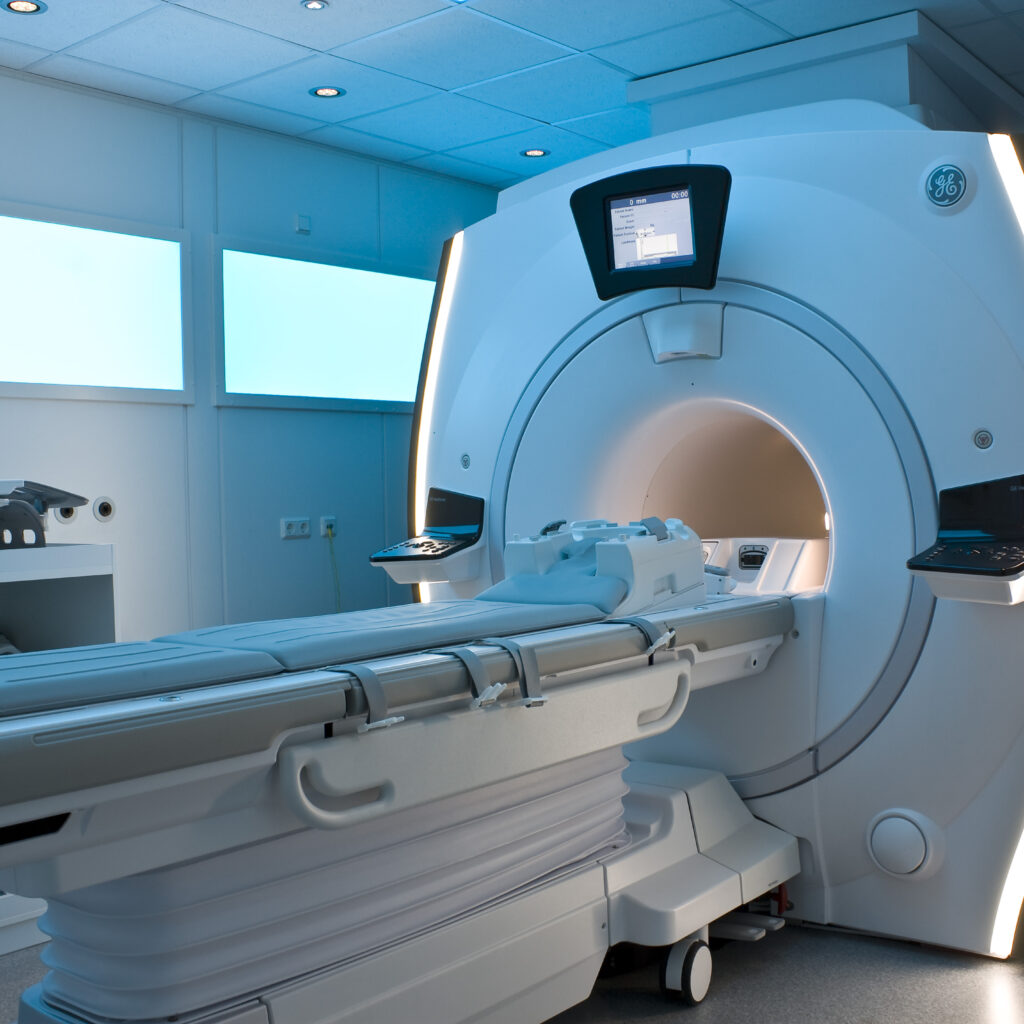
Magnetic Resonance Imaging (MRI) is a cutting-edge medical imaging technique that offers detailed visualization of the internal structures of the human body. It is widely used to generate high-resolution images of organs, soft tissues, and neural pathways, aiding in diagnosis, treatment planning, and ongoing patient monitoring. MRI’s non-invasive nature and ability to differentiate between various tissue types make it a cornerstone in modern medical diagnostics.
Fundamental Principles Behind MRI
MRI operates by harnessing the magnetic properties of atomic nuclei, predominantly hydrogen atoms, which are abundant in the body due to its high water content. When a patient is placed inside a strong magnetic field generated by the MRI machine, the hydrogen nuclei align with this field. The system then emits radiofrequency (RF) pulses that temporarily disturb this alignment. As the nuclei return to their original state, they emit signals that are detected by the scanner. These signals are processed using advanced computational algorithms to produce detailed images of the body’s internal structures.
- Hydrogen Nuclei Alignment:
The strong magnetic field causes hydrogen protons to align along the field lines. - Radiofrequency Excitation:
RF pulses disrupt this alignment, causing nuclei to absorb energy. - Signal Emission and Detection:
As nuclei relax back, they emit signals captured by the scanner. - Image Reconstruction:
Complex algorithms convert these signals into detailed anatomical images.
Types of MRI Techniques
MRI technology encompasses various specialized scanning methods, each designed to highlight different tissue characteristics or physiological functions.
Standard MRI Scans
Traditional MRI scans primarily use T1-weighted and T2-weighted sequences. T1-weighted images excel at depicting anatomical details, especially tissues rich in fat, while T2-weighted images are more sensitive to fluids, making them ideal for detecting inflammation, edema, or other fluid-related abnormalities.
Functional MRI (fMRI)
Functional MRI measures brain activity by detecting changes in blood flow and oxygenation. This technique maps active brain regions during specific tasks, providing critical information for neurosurgical planning and cognitive research by identifying areas responsible for speech, movement, and other vital functions.
Diffusion Tensor Imaging (DTI)
DTI is a specialized MRI method that visualizes the brain’s white matter tracts by tracking the diffusion of water molecules along neural pathways. This imaging is essential for assessing the integrity of neural connections, particularly in cases of neurodegenerative diseases or brain injuries.
Magnetic Resonance Angiography (MRA)
MRA focuses on imaging blood vessels non-invasively, using MRI principles to create detailed pictures of arteries and veins. It is instrumental in detecting vascular conditions such as aneurysms or stenosis, often without the need for contrast agents, although contrast-enhanced MRA can improve image clarity.
Clinical Applications of MRI
MRI’s versatility allows it to serve multiple medical disciplines by providing critical diagnostic information.
Neurological Uses
MRI is the preferred imaging modality for diagnosing brain and spinal cord disorders, including tumors, strokes, multiple sclerosis, and degenerative diseases. Its high-resolution images enable precise localization and characterization of neurological abnormalities.
Orthopedic Applications
In orthopedics, MRI is invaluable for evaluating soft tissue injuries, joint pathologies, and musculoskeletal tumors. It offers detailed visualization of cartilage, ligaments, and tendons, making it essential in sports medicine and rehabilitation.
Oncological Role
MRI assists oncologists in detecting tumors, determining their stage, and monitoring treatment response. It provides detailed information about tumor size, location, and involvement of surrounding tissues, which is crucial for effective treatment planning.
Advantages of MRI
- Absence of Ionizing Radiation:
MRI uses magnetic fields and radio waves instead of harmful ionizing radiation, making it safer for repeated use. - Superior Image Resolution:
It delivers high-contrast images that differentiate soft tissues with exceptional clarity. - Wide Range of Applications:
Various MRI techniques can be tailored to specific diagnostic needs across multiple medical fields.
Limitations and Challenges of MRI
- High Cost:
MRI scans are often expensive, which can limit accessibility for some patients. - Lengthy Procedure Times:
The scanning process can be time-consuming, requiring patients to remain still, which may be difficult for certain individuals. - Contraindications with Metal Implants:
Patients with devices like pacemakers or certain implants may be unable to undergo MRI due to safety concerns.
Why MRI is Essential in Modern Medicine
MRI has transformed medical imaging by providing detailed, non-invasive insights into the body’s internal structures and functions. Its ability to produce high-quality images without radiation exposure makes it indispensable for diagnosis, treatment planning, and monitoring across numerous medical specialties. Understanding MRI’s principles, applications, and limitations empowers healthcare providers and patients to make informed decisions, ultimately improving clinical outcomes.
FAQ
What is the principle behind MRI?
MRI exploits the magnetic properties of hydrogen nuclei in the body aligned by a strong magnetic field and excited by radiofrequency pulses to produce images.
What distinguishes T1-weighted and T2-weighted MRI scans?
T1-weighted images highlight fat content and anatomical detail, while T2-weighted images emphasize fluid-filled areas such as inflammation or edema.
What clinical conditions can MRI help diagnose?
MRI is used for detecting tumors, strokes, neurodegenerative diseases, soft tissue injuries, and vascular abnormalities.
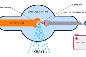
Why is MRI preferred over X-rays or CT in some cases?
Because MRI does not use ionizing radiation and provides superior soft tissue contrast, making it safer and more effective for certain diagnostics.
Can MRI be used with metal implants?
Certain metal implants like pacemakers can pose risks, so MRI may be contraindicated depending on the implant type.


Leave a Reply