Short Answer
Definition of Molecular Imaging
Molecular imaging is a cutting-edge technique in biomedical science and clinical medicine that enables the visualization, characterization, and measurement of biological processes at the molecular and cellular levels within living organisms. Unlike traditional imaging methods that primarily depict anatomical structures, molecular imaging focuses on the dynamic biochemical activities underlying health and disease, providing critical insights into cellular function and molecular pathways.
- Scope:
It encompasses a variety of imaging modalities designed to detect specific molecular targets or physiological changes. - Purpose:
To enhance diagnosis, monitor therapeutic responses, and facilitate personalized treatment strategies by revealing molecular alterations before anatomical changes become apparent.
Key Modalities of Molecular Imaging
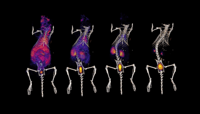
Positron Emission Tomography (PET)
Positron Emission Tomography is a non-invasive imaging technique that employs radiotracers labeled with positron-emitting isotopes. When these isotopes decay, they emit positrons that annihilate with electrons, producing pairs of gamma photons detected by the PET scanner. This process allows real-time visualization of metabolic and physiological functions.
- Applications:
PET is extensively used in oncology to detect tumors by tracking increased glucose metabolism using fluorodeoxyglucose (FDG), in neurology for brain function studies, and in cardiology to assess myocardial viability. - Advantages:
High sensitivity to metabolic changes and the ability to quantify biochemical processes.
Single Photon Emission Computed Tomography (SPECT)
SPECT imaging operates on a principle akin to PET but utilizes gamma-emitting radioisotopes detected by gamma cameras. Although it generally offers lower spatial resolution than PET, SPECT excels in functional imaging, particularly in cardiac assessments.
- Clinical Use:
Commonly used to evaluate myocardial perfusion and diagnose coronary artery disease using agents like technetium-99m. - Benefits:
More accessible and cost-effective compared to PET, making it widely available in clinical practice.
Magnetic Resonance Imaging (MRI) in Molecular Imaging
While MRI is traditionally known for its high-resolution anatomical imaging, advancements have expanded its role into molecular imaging through specialized contrast agents and techniques such as Magnetic Resonance Spectroscopy (MRS). These innovations enable the assessment of metabolic and molecular changes within tissues.
- Techniques:
Use of hyperpolarized agents to track metabolic pathways in vivo. - Significance:
Provides exceptional soft tissue contrast and spatial resolution without ionizing radiation.
Optical Imaging Techniques
Optical imaging encompasses methods like bioluminescence and fluorescence imaging, which detect light emitted from genetically modified proteins or fluorescent dyes introduced into biological systems. This modality is highly sensitive and versatile, particularly suited for small animal research.
- Applications:
Real-time monitoring of molecular events and disease progression in preclinical models. - Limitations:
Limited tissue penetration restricts its use in larger animals and humans, prompting ongoing research to enhance depth capabilities.
Theranostics: Combining Therapy and Diagnostics
Theranostics represents an innovative approach that integrates molecular imaging with targeted therapy. This strategy employs radiopharmaceuticals designed to both visualize and treat disease simultaneously, enabling personalized medicine tailored to individual patient profiles.
- Example:
In prostate cancer, agents such as ^223Radium are used to detect and treat metastatic lesions concurrently. - Impact:
Enhances treatment precision and monitoring, improving clinical outcomes.
Computed Tomography (CT) in Hybrid Imaging
Although primarily a structural imaging tool, CT is frequently combined with molecular imaging modalities to provide comprehensive diagnostic information. Hybrid systems like PET/CT merge metabolic data from PET with anatomical detail from CT scans.
- Benefit:
Enables precise localization of molecular abnormalities within anatomical context, crucial for accurate diagnosis and intervention planning. - Clinical Relevance:
Widely used in oncology, cardiology, and neurology for enhanced diagnostic accuracy.
Emerging Advances: Nanoimaging
Nanoimaging leverages nanoparticles engineered with unique optical and magnetic properties to serve as highly sensitive and specific contrast agents. This frontier in molecular imaging holds promise for early disease detection and targeted drug delivery.
- Capabilities:
Nanoparticles can identify pathological changes at very early stages, often before symptoms arise. - Therapeutic Potential:
Facilitate precise delivery of therapeutic agents, minimizing side effects and maximizing efficacy.
How Molecular Imaging Works
Molecular imaging techniques function by introducing specific probes or contrast agents that interact with targeted molecules or cellular processes. These interactions produce detectable signals-such as gamma rays, magnetic resonance changes, or light emissions-that are captured by specialized imaging devices. The resulting images reveal the spatial distribution and intensity of molecular activity, enabling detailed analysis of physiological and pathological states.
Importance of Molecular Imaging
The significance of molecular imaging lies in its ability to transform medical diagnostics and treatment by providing a window into the molecular underpinnings of disease. It facilitates early detection, accurate diagnosis, and real-time monitoring of therapeutic responses, thereby enhancing patient management and outcomes. Furthermore, molecular imaging accelerates biomedical research by elucidating complex biological mechanisms, fostering the development of novel therapies.
Common Misconceptions About Molecular Imaging
Molecular imaging only provides anatomical information.
Unlike traditional imaging, molecular imaging reveals functional and biochemical processes, not just structural details.
All molecular imaging techniques involve high radiation exposure.
While some modalities like PET and SPECT use radioactive tracers, others such as MRI and optical imaging do not involve ionizing radiation.
Optical imaging is suitable for human clinical use.
Due to limited tissue penetration, optical imaging is primarily used in small animal research, with ongoing efforts to adapt it for clinical applications.
Real-World Applications of Molecular Imaging
Molecular imaging is extensively applied across various medical fields:
- Oncology:
Detecting and staging cancers, monitoring treatment response, and guiding targeted therapies. - Neurology:
Investigating brain metabolism, receptor binding, and neurodegenerative diseases. - Cardiology:
Assessing myocardial perfusion, viability, and detecting coronary artery disease. - Drug Development:
Evaluating pharmacodynamics and pharmacokinetics of new therapeutics in vivo.
FAQ
What is molecular imaging?
Molecular imaging is a biomedical technique that visualizes, characterizes, and measures biological processes at the molecular and cellular levels within living organisms.
What are the main types of molecular imaging?
Main types include Positron Emission Tomography (PET), Single Photon Emission Computed Tomography (SPECT), Magnetic Resonance Imaging (MRI) with molecular techniques, optical imaging, theranostics, and nanoimaging.
How does molecular imaging differ from traditional imaging?
Unlike traditional imaging that shows anatomical structures, molecular imaging reveals functional and biochemical processes, enabling earlier disease detection and personalized treatment.
Is molecular imaging safe in terms of radiation exposure?
Some molecular imaging techniques like PET and SPECT involve radiation, but others such as MRI and optical imaging do not use ionizing radiation.
What are the clinical applications of molecular imaging?
It is used in oncology, neurology, cardiology, and drug development to detect diseases, monitor therapy, and guide personalized treatments.


Leave a Reply