Short Answer
Definition
The use of electron beams to visualize DNA represents a groundbreaking approach at the crossroads of quantum physics and molecular biology. This technique enables detailed imaging of biological macromolecules, particularly deoxyribonucleic acid (DNA), without causing damage to the samples. Unlike traditional methods that may compromise the structural integrity of DNA, electron-based imaging offers a non-destructive alternative that preserves the native state of genetic material, facilitating more accurate analysis.
Principles of Electron-Based DNA Imaging
Electron microscopy leverages the dual wave-particle nature of electrons to achieve resolution far beyond that of conventional light microscopy. When electrons interact with matter, their short wavelengths allow for the detection of atomic-scale features, making them ideal for examining the fine structural details of DNA molecules. This high-resolution capability is essential for studying sub-cellular components where precision and clarity are critical.
Types of Electron Microscopy Techniques
- Scanning Electron Microscopy (SEM):
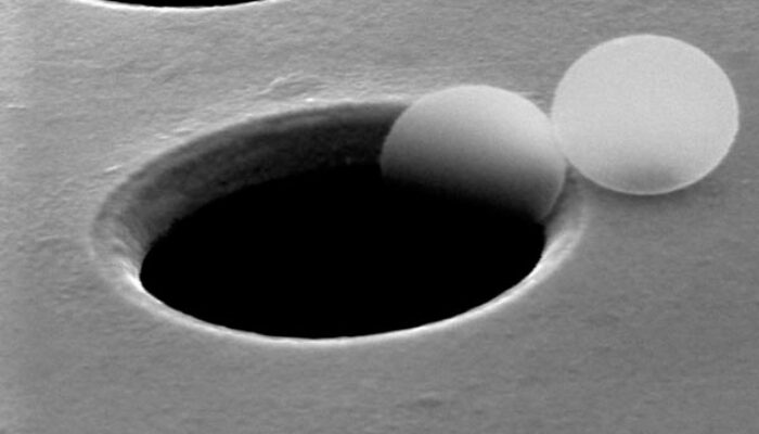
SEM generates three-dimensional images by scanning the surface of DNA samples with a focused electron beam, revealing topographical details. - Transmission Electron Microscopy (TEM):
TEM transmits electrons through ultra-thin DNA specimens, providing detailed internal structural views at the molecular level. - Cryo-Electron Microscopy (Cryo-EM):
Cryo-EM involves rapid freezing of DNA samples at cryogenic temperatures, preserving their native conformation and minimizing artifacts caused by dehydration or chemical fixation.
Sample Preparation and Preservation
Preparing DNA samples for electron microscopy is a delicate process that significantly influences imaging outcomes. Traditional preparation methods-such as chemical fixation, dehydration, and embedding-can alter or damage DNA structures. Cryo-EM has revolutionized this step by flash-freezing samples, which maintains the DNA’s natural state and secondary structures, allowing for more authentic visualization.
Structural Insights Enabled by Electron Imaging
Electron microscopy provides unparalleled detail in examining DNA architecture. It allows researchers to observe nuclear components like chromatin organization and nucleosome positioning with exceptional clarity. This technology also facilitates the study of DNA conformational changes under varying environmental conditions, including temperature shifts, ionic concentrations, and interactions with DNA-binding proteins. Such insights deepen our understanding of essential biological processes such as DNA replication, transcription, and repair.
Dynamic Visualization of DNA Processes
Advanced electron microscopy techniques enable real-time observation of DNA dynamics, offering valuable perspectives on molecular events. For instance, the assembly and disassembly of chromatin structures-key to gene regulation-can be monitored as they occur. This temporal resolution captures transient molecular states that static imaging methods often miss, providing a more comprehensive view of genetic function and regulation.
Advantages of Non-Destructive Electron Imaging
One of the most significant benefits of electron-based DNA imaging is its minimally invasive nature. This allows for repeated examination of the same sample over time, facilitating longitudinal studies that track DNA changes in response to various stimuli or treatments. Such capability is particularly beneficial in pharmaceutical research, where monitoring the effects of drugs on genetic material is crucial for developing effective therapies.
Integration of Computational Technologies
The fusion of electron microscopy with machine learning and artificial intelligence has markedly enhanced data processing and interpretation. Automated algorithms can analyze vast datasets rapidly, identifying subtle patterns and structural features that might elude human observers. This synergy between high-resolution imaging and computational analysis is transforming biological research by enabling more precise and comprehensive understanding of DNA structures.
Challenges and Limitations
- Technical Complexity:
Operating electron microscopes requires specialized skills in instrumentation, sample preparation, and data interpretation, posing a barrier to widespread adoption. - Resource Intensity:
Techniques like cryo-EM demand expensive equipment and controlled environments, limiting accessibility for some research facilities. - Sample Integrity:
Despite advances, maintaining completely unaltered DNA samples remains challenging, necessitating ongoing refinement of protocols.
Ethical Considerations
As electron microscopy unveils intricate details of DNA, ethical issues related to genetic privacy, manipulation, and bioethical standards come to the forefront. Responsible application of these powerful imaging tools is essential to ensure that scientific progress aligns with societal values and safeguards individual rights.
Significance and Future Prospects
The capability to image DNA using electrons epitomizes a remarkable fusion of cutting-edge technology and molecular biology. This non-destructive approach unlocks new research possibilities, offering unprecedented insights into the molecular underpinnings of life. Continued advancements promise to revolutionize biotechnology, medicine, and genetic research, pushing the boundaries of scientific exploration and deepening our comprehension of genetic material at the most fundamental level.
FAQ
What is the main advantage of using electron microscopy for DNA imaging?
The main advantage is its non-destructive nature, allowing for detailed imaging of DNA without compromising its structural integrity.
What types of electron microscopy are used for DNA analysis?
The main types include Scanning Electron Microscopy (SEM), Transmission Electron Microscopy (TEM), and Cryo-Electron Microscopy (Cryo-EM).
What challenges are associated with electron microscopy?
Challenges include the technical complexity of operation, resource intensity of certain techniques, and difficulties in maintaining sample integrity.


Leave a Reply