Short Answer
Definition of Diagnostic Medical Sonography
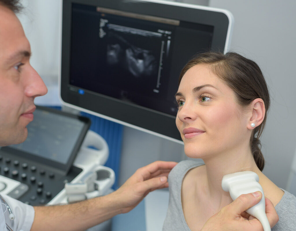
Diagnostic medical sonography, commonly known as ultrasound imaging, is a pivotal medical technology that merges sound wave physics with clinical diagnostics. It enables healthcare professionals to visualize internal body structures in real-time without invasive procedures. This imaging technique is widely utilized across numerous medical specialties to assist in both diagnosis and treatment.
Fundamental Principles of Ultrasound Imaging
At its essence, sonography uses high-frequency sound waves generated by a handheld device called a transducer. These sound waves travel through body tissues and reflect back when they encounter different structures. The returning echoes are captured and converted into visual images, allowing clinicians to observe organs, tissues, and blood flow. Unlike X-rays or CT scans, ultrasound does not involve ionizing radiation, making it a safer option for repeated use.
Applications Across Medical Specialties
Obstetrics and Prenatal Care
Ultrasound plays a crucial role in monitoring fetal development during pregnancy. It provides detailed images of the fetus inside the womb, helping assess growth, detect abnormalities, and evaluate overall health. This non-invasive method offers expectant parents and clinicians valuable insights into early life stages.
Cardiology and Heart Evaluation
In cardiology, sonography is employed through echocardiography to examine the heart’s anatomy and function. It visualizes heart chambers, valves, and blood flow patterns, aiding in the diagnosis of cardiac diseases. Doppler ultrasound further enhances this by measuring the velocity and direction of blood flow, offering critical information about cardiovascular health.
Abdominal and Organ Imaging
Sonography is extensively used to inspect abdominal organs such as the liver, kidneys, gallbladder, and pancreas. It helps detect conditions like gallstones, liver cirrhosis, kidney stones, and tumors. Additionally, ultrasound guides procedures like biopsies and monitors disease progression, providing a non-invasive alternative to exploratory surgery.
Technical Aspects and Image Formation
The transducer acts as both an emitter and receiver of ultrasound waves. When sound waves encounter tissues of varying densities, they reflect differently-a property known as echogenicity. Dense structures like bone and air produce strong echoes and appear bright on the image, whereas fluid-filled areas appear dark due to low reflection. This contrast enables detailed visualization of anatomical features.
Challenges in Image Interpretation
Interpreting ultrasound images requires significant expertise. The quality of the images can be affected by patient-specific factors such as body composition and anatomical variations. Artifacts-unintended echoes or shadows-may obscure true structures, complicating diagnosis. Therefore, the sonographer’s skill and experience are critical in distinguishing between normal and pathological findings.
Limitations of Ultrasound Technology
- Bone and Gas Interference:
Ultrasound waves cannot effectively penetrate bones or gas-filled organs, limiting visualization of certain areas like the brain or lungs. - Image Quality in Obesity:
Increased tissue thickness in obese patients can degrade image clarity, sometimes necessitating complementary imaging techniques.
Advancements in Sonographic Techniques
Recent technological progress has introduced three-dimensional (3D) and four-dimensional (4D) ultrasound imaging. These innovations provide volumetric and real-time dynamic views of anatomical structures, enhancing diagnostic precision. For example, 3D ultrasound offers detailed fetal morphology, while 4D adds the element of motion, improving assessment of organ function and development.
Importance of Training and Interdisciplinary Collaboration
As ultrasound technology evolves, continuous education for sonographers is essential to maintain high diagnostic standards. Skilled professionals must adapt to new tools and techniques to maximize patient outcomes. Collaboration among radiologists, obstetricians, cardiologists, and other specialists ensures comprehensive interpretation and integrated care.
Significance of Diagnostic Medical Sonography
Diagnostic medical sonography is a cornerstone of modern healthcare, providing a safe, versatile, and cost-effective imaging solution. Its ability to deliver real-time insights into the human body without radiation exposure makes it indispensable in clinical practice. As technology and expertise advance, ultrasound will continue to expand its role in unraveling complex medical conditions and guiding therapeutic interventions.
FAQ
What is diagnostic medical sonography?
Diagnostic medical sonography is a non-invasive imaging technique that uses high-frequency sound waves to visualize internal body structures in real-time for medical diagnosis.
Is ultrasound imaging safe?
Yes, ultrasound does not use ionizing radiation, making it a safer imaging option compared to X-rays or CT scans, especially for repeated use.
What medical fields use sonography?
Sonography is widely used in obstetrics, cardiology, abdominal imaging, and many other medical specialties for diagnosis and treatment guidance.
What are the limitations of ultrasound imaging?
Ultrasound waves cannot effectively penetrate bones or gas-filled organs, and image quality can be reduced in patients with obesity.
What recent advancements exist in sonographic technology?
Recent advancements include 3D and 4D ultrasound imaging, which provide volumetric and real-time dynamic views for enhanced diagnostic precision.


Leave a Reply