In the realm of medical imaging, the term “radiology image” encompasses a variety of intricate imaging modalities and techniques that physicians and radiologists utilize to diagnose and manage medical conditions. At its core, a radiology image serves as a visual representation of the internal structures and functions of the body, produced through various methods. The technical term for a radiology image often varies based on the imaging technique employed. Common terms include “X-ray,” “computed tomography (CT) scan,” “magnetic resonance imaging (MRI),” “ultrasound,” and “nuclear medicine scans.” Each type of imaging technique offers unique insights and diagnostic capabilities that are crucial in contemporary medical practice.
First, let us explore the ubiquitous X-ray, perhaps the most recognized form of radiology imagery. X-rays harness high-frequency electromagnetic radiation to penetrate through the body, capturing images of dense structures such as bones. The technical term for an X-ray image is often simply “X-ray film” or “radiograph.” This imaging modality is indispensable in emergency settings, where rapid assessment of fractures, dislocations, and foreign bodies can drastically alter patient management. In advanced applications, digital X-ray techniques enhance image quality and reduce radiation exposure, highlighting the evolution of this traditional technique.
Next, the computed tomography (CT) scan provides a more comprehensive visual narrative. A CT scan amalgamates multiple X-ray images taken from various angles, employing computer algorithms to produce cross-sectional images or “slices” of the body. These images allow for an unparalleled visualization of soft tissues, organs, and blood vessels, significantly aiding the diagnosis of complex conditions such as tumors, internal bleeding, or infectious lesions. The terminology associated with CT imaging includes “CT scan” or “computed axial tomography (CAT) scan,” emphasizing its sophisticated technology. Additionally, the advent of “3D CT” reconstruction enables a more detailed understanding of anatomical structures, heralding a new era in diagnostic imaging.
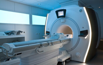
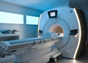
Magnetic resonance imaging (MRI) represents another pivotal component of radiological evaluation, employing magnetic fields and radio waves to produce detailed images of soft tissues. The technical terminology for an MRI image typically includes “MRI scan” or “MR image.” MRI is particularly adept at visualizing neural tissues and musculoskeletal structures, thus proving invaluable in diagnosing neurological disorders, joint abnormalities, and soft tissue injuries. Importantly, MR imaging is well-regarded for its ability to provide high-resolution images without exposing patients to ionizing radiation, a significant advantage over X-rays and CT scans.
As we delve deeper into sonography, or ultrasound imaging, the technical term shifts to “ultrasonic image” or simply “ultrasound.” This modality employs high-frequency sound waves to generate real-time images, making it indispensable for obstetric evaluations, cardiac assessments, and abdominal examinations. Ultrasound imaging is particularly advantageous due to its portability, ability to manipulate images in real time, and lack of ionizing radiation. It is commonly used in clinical environments to visualize organs, assess blood flow, and guide interventional procedures, showcasing its versatility in medical contexts.
The domain of nuclear medicine introduces a distinctive array of imaging techniques involving the administration of radiopharmaceuticals. The technical nomenclature includes “nuclear scan” or “radiologic scintigraphy.” These imaging modalities provide functional data about organs and tissues by observing the distribution of radioactive tracers within the body. Nuclear scans are particularly effective in evaluating conditions such as thyroid dysfunction, cardiac perfusion, and the detection of malignancies. The use of positron emission tomography (PET) in conjunction with CT scans has heralded a new era in oncology, where both anatomical and functional information simultaneously allows for precise staging and response assessment in cancer patients.
Furthermore, one must consider the implications of hybrid imaging technologies, which amalgamate multiple imaging modalities for enhanced diagnostic capabilities. Techniques like PET/CT and SPECT/CT epitomize advancements in radiation imaging, wherein the complementary nature of functional and anatomical assessment may provide a more holistic view of disease processes. The terminology associated with such hybrid modalities includes “fusion imaging” or “integrated imaging,” which underscores their potential to significantly improve diagnostic accuracy and therapeutic planning.
In summary, the universe of radiology images encompasses a diverse array of modalities, each with its own defining technical terminology, mechanisms of action, and clinical applications. From traditional X-rays to advanced MRI techniques and innovative nuclear imaging approaches, radiology images serve as critical tools in the diagnostic arsenal of modern medicine. Understanding the specific terms associated with each modality enriches the dialogue between healthcare practitioners and patients, fostering a nuanced comprehension of the diagnostic processes at play. These images not only illuminate anatomical structures but also delineate the pathways of physiological functions, thus enabling healthcare professionals to deliver personalized, effective, and timely medical care.