Magnetic Resonance Imaging (MRI) is a sophisticated imaging technique that has revolutionized the field of medicine, particularly in diagnostics and treatment planning. It offers profound insights into the human body’s internal structures without the use of ionizing radiation, distinguishing itself from other imaging modalities like X-rays and computed tomography (CT) scans. MRI has become an indispensable tool in various specialties, including neurology, orthopedics, and oncology, as it provides unparalleled detail of soft tissues, which often elude other imaging techniques.
At the core of MRI technology lies the principle of nuclear magnetic resonance (NMR), a phenomenon that exploits the magnetic properties of certain atomic nuclei. When placed in a strong magnetic field, nuclei of atoms, particularly hydrogen which is abundant in the human body, resonate at a frequency determined by their atomic environment. This resonance allows for the precise manipulation and measurement of the atoms, culminating in the creation of highly detailed images. The significance of MRI is not just limited to visualizing anatomical structures; its capacity to differentiate between various tissue types has paved the way for advanced diagnostic capabilities.
Principle Mechanism of MRI
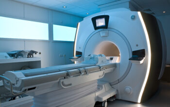
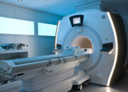
The operational mechanics of MRI encompass several key stages: the magnetic field generation, radiofrequency pulse application, and data acquisition. Initially, the patient is situated within a powerful superconducting magnet that generates a magnetic field typically ranging from 1.5 to 3 Tesla. This magnetic field aligns the protons in the hydrogen nuclei of water molecules in the body. When the protons are oriented with the magnetic field, they are in a state of equilibrium.
Upon the application of a radiofrequency (RF) pulse, a brief yet potent burst of electromagnetic energy, the protons are momentarily displaced from their equilibrium state. This disruption causes a phenomenon known as resonance, where protons absorb energy and shift to a higher energy state. Moreover, when the RF pulse is discontinued, the protons slowly return to their original alignment, releasing energy in the form of signals that can be detected and measured by the MRI scanner’s receiver coils.
Image Formation
The energies emitted by the returning protons vary based on their surrounding environments and the types of tissues they occupy. This variability in relaxation times allows MRI software to construct detailed images that reflect the distinct characteristics of various tissues. Two primary relaxation processes are instrumental in this data collection: T1 (spin-lattice) and T2 (spin-spin) relaxation times. T1 relates to the time it takes for protons to return to equilibrium, while T2 pertains to the time it takes for protons to lose phase coherence among neighboring spins.
Different sequences of RF pulses and magnetic gradients can be employed to accentuate these relaxation characteristics, yielding diverse imaging modalities such as T1-weighted, T2-weighted, and diffusion-weighted imaging (DWI). Each modality has its unique strengths, allowing radiologists to tailor MRI scans to best visualize specific pathological conditions, enhancing the diagnostic accuracy fundamentally.
Applications of MRI
The versatility of MRI is evidenced by its myriad of applications across various clinical fields. In neurology, MRI is the gold standard for detecting brain tumors, assessing demyelinating diseases such as multiple sclerosis, and evaluating acute stroke scenarios where time is of the essence. The exquisite detail of brain structures allows clinicians to devise appropriate management strategies for various neurologic diseases.
Within orthopedics, MRI serves a critical role in evaluating musculoskeletal disorders. It provides unparalleled insight into soft tissue injuries—such as ligament tears and cartilage damage—commonly occurring in athletes. The ability to visualize these structures helps in devising rehabilitation protocols, and in some cases, surgical planning, highlighting its importance in sports medicine.
Moreover, the oncology realm has increasingly adopted MRI for tumor characterization and treatment monitoring. It aids in assessing tumor vascularity and response to therapies, thus playing a pivotal role in personalized medicine initiatives. Functional MRI (fMRI) has also pioneered non-invasive exploration of brain activity. By measuring changes in blood flow, fMRI can elucidate neural pathways involved in specific tasks, bridging the gap between structure and function.
Ethical Considerations and Future Prospects
While MRI is heralded for its non-invasive and extraordinarily detailed imaging capabilities, it is not without its challenges and ethical considerations. The necessity for metal-free environments to ensure patient safety during scanning is paramount, especially for individuals with pacemakers or certain implants. Moreover, the accessibility of MRI can be constrained by high operational costs and the need for specialized technicians and radiologists.
Looking ahead, ongoing advancements in MRI technology are poised to enhance its application further. Innovations such as diffusion tensor imaging (DTI) allow for the visualization of white matter tracts in the brain, while advances in imaging software are making scans more rapid and less cumbersome for patients. Research into a variety of contrast agents also aims to enhance the specificity and sensitivity of MRI, fostering better detection of malignancies and other conditions at earlier stages.
In conclusion, MRI scanning embodies a fascinating intersection of physics, biology, and medical science, facilitating an array of clinical applications that significantly contribute to modern healthcare. Its ability to unveil the intricacies of the human body without invasive procedures continues to invoke interest and respect in medical communities and among patients alike. The ongoing evolution of MRI technology promises an even brighter future, heralding unparalleled diagnostic capabilities while continuing to uphold patient safety and tolerability as fundamental principles.