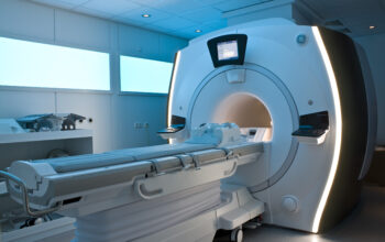
Magnetic Resonance Imaging (MRI) transcends the conventional realm of medical imaging, embodying a sophisticated interplay between physics and biology. Its allure lies not only in its advanced technology but also in its ability to unveil the enigmatic intricacies of the human body, akin to an artist peeling back layers of a masterpiece to reveal the vibrant details beneath. This essay delves into the fundamental principles, operational mechanisms, clinical applications, and future prospects of MRI, offering an expansive vista into this remarkable diagnostic tool.
To comprehend the essence of MRI, it is essential to first grasp the underlying physics that powers this modality. At its core, MRI utilizes the principles of nuclear magnetic resonance (NMR), where certain atomic nuclei exhibit magnetic properties when exposed to an external magnetic field. The most prevalent nuclei observed in the human body are hydrogen nuclei, primarily sourced from water molecules. Water constitutes approximately 70% of our body, making hydrogen a plentiful and instrumental target for imaging.
In the presence of a strong magnetic field, typically ranging from 1.5 to 3.0 teslas, the nuclear spins of hydrogen nuclei align with the field. A radiofrequency pulse is then introduced, which excites these spins, prompting them to resonate. Once the pulse is halted, the nuclei return to their ground state, emitting energy in the process. This emitted energy is detected by the MRI scanner and transformed into a visual representation of the internal structures. The magnetic resonance phenomenon is analogous to a finely tuned musical instrument, where the correct frequencies yield a harmonious image.
The intricacies of MRI extend beyond mere image acquisition; they encompass nuances that contribute to the quality and contrast of the generated images. Different tissues within the human body respond uniquely to the magnetic fields and radiofrequency pulses, leading to varying relaxation times. Two critical parameters in this regard are T1 (spin-lattice) and T2 (spin-spin) relaxation times. T1 relaxation relates to the time taken for hydrogen nuclei to realign with the magnetic field after being disturbed, while T2 relaxation pertains to the time taken for the spins of neighboring nuclei to de-phase. These differing relaxation times allow for the contrast between various tissues, providing radiologists with enhanced clarity to distinguish between healthy and pathological states.
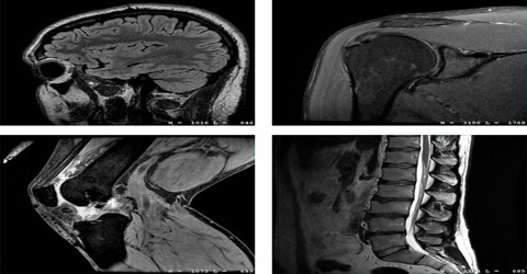
An invaluable facet of MRI is its multifaceted clinical applications. This technique has emerged as a cornerstone in diagnosing a plethora of conditions across numerous medical specialties. In the realm of neurology, MRI is instrumental in visualizing brain structures and identifying anomalies such as tumors, strokes, or degenerative diseases. The clarity with which MRI delineates the gray and white matter creates a vivid tapestry of neurological architecture, often revealing conditions that other imaging modalities cannot.
Moreover, the orthopedic domain thrives on the versatility of MRI, adept at assessing soft tissue injuries, cartilage deterioration, and ligament tears. Unlike X-rays, which predominantly exhibit bony structures, MRI’s capacity to render soft tissue details brings to light injuries often obscured by osseous shadows. The images generated possess an ethereal quality, almost reminiscent of a ghostly reflection of anatomical structures, allowing clinicians to comprehend the complexities of musculoskeletal injuries.
Furthermore, MRI finds its applications in the field of oncology for tumor detection, staging, and treatment monitoring. The imaging nuances facilitate the observation of malignancies in various tissues, allowing for targeted therapeutic interventions. The contrast agents utilized, such as gadolinium-based compounds, further enhance the visualization of particular lesions, akin to an artist accentuating vibrant colors against a subtle background.
Beyond traditional applications, MRI continuously evolves, carving pathways for innovative techniques that enhance its diagnostic prowess. Functional MRI (fMRI), for instance, has emerged as a revolutionary advancement, enabling researchers to map brain activity by detecting changes in blood flow. This dynamic imaging technique unveils the brain’s electrophysiological patterns during cognitive tasks, illuminating the intricacies of functional connectivity and neuropathology.
Additionally, developments in diffusion-weighted imaging (DWI) allow for the visual assessment of water molecule diffusion within tissues, rendering insights into cellular integrity and cellular density. This modality proves particularly beneficial in detecting early stroke signs, heralding a potential paradigm shift in emergency medicine.
As MRI technology continues to advance, the horizon appears boundless. Future innovations may well encompass ultra-high-field MRI scanners, providing unprecedented resolution and specificity. Quantum technologies may revolutionize coil design, amplifying signal detection and heralding a new era in imaging clarity. Moreover, the integration of artificial intelligence into MRI analysis has the potential to expedite diagnosis significantly, rendering a once time-consuming endeavor into a swift, precise process.
In conclusion, Magnetic Resonance Imaging stands as a testament to the convergence of physics and medicine, illuminating the intricacies of human anatomy with unparalleled sophistication and detail. Its operational foundations, diverse applications, and continuous technological advancements coalesce to maintain its position as an indispensable tool within the diagnostic arsenal. By navigating through the complexities of MRI, one glimpses an art that marries science and empathy—a truly remarkable achievement in the annals of medical history. As we look to the future, the promise of MRI continues to inspire, presenting an exquisite canvas upon which the stories of our health are painted.