In the realm of modern medicine, computed tomography (CT) scans stand as a pinnacle of diagnostic prowess, offering intricate cross-sectional imagery indispensable for accurate diagnosis and treatment planning. However, with the increasing utilization of this technology, questions arise regarding the implications of repeated exposure to ionizing radiation. This article investigates the delicate equilibrium between the advantages of CT imaging and the compounded risks associated with multiple scans, delving into the underlying physics that governs radiation exposure in medical imaging.
Understanding Ionizing Radiation
At the crux of the discourse on CT scans and their risks lies the concept of ionizing radiation. Ionizing radiation is capable of displacing electrons from atoms, leading to the formation of charged particles or ions. In the context of medical imaging, this type of radiation can be beneficial, as it enables the production of detailed images due to differential attenuation of X-rays by various tissues. However, the detrimental effects of ionizing radiation, particularly at high doses, must not be underestimated.
When discussing the safety thresholds associated with CT scans, it is crucial to understand the concept of dose measurement. The effective dose from a CT scan is quantified in sieverts (Sv), a unit that accounts for the biological effects of radiation on human tissue. The average effective dose from a standard CT scan of the abdomen is approximately 10 mSv, which is significantly higher than the annual natural background radiation exposure (about 2-3 mSv). Understanding these metrics is essential for comprehending how cumulative doses can escalate with repeated scans.
Cumulative Exposure: A Quantitative Approach
One of the primary concerns surrounding repeated CT scans involves cumulative exposure to radiation. The linear no-threshold (LNT) model posits that there is a proportional relationship between radiation dose and the risk of developing cancer; even small doses of radiation are considered to carry a risk. This model suggests that the risk of radiation-induced malignancies escalates with each subsequent scan. For instance, a patient receiving multiple abdominal CT scans over a short duration may accumulate an effective dose that substantially raises their lifetime risk of exposure-related cancers.
Research indicates that the risk of a fatal cancer from a 10 mSv exposure is approximately 1 in 2,000. While these numbers are contingent upon numerous variables—such as age at exposure, sex, and genetic factors—this statistic highlights the compelling need for justifiable indications before performing repetitive imaging.
Risk-Benefit Analysis of CT Scans
The primary benefit of CT scanning lies in its unparalleled ability to reveal internal tissue structures with remarkable clarity, aiding in the diagnosis of complex conditions—ranging from acute trauma to oncology. However, it is vital for healthcare providers to engage in a thorough risk-benefit analysis when determining the necessity of a CT scan. This includes evaluating clinical indications vis-à-vis the associated radiation risks.
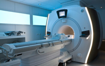
In cases such as acute abdominal pain, where quick diagnosis is critical, the expediency of a CT scan may outweigh potential hazards. Conversely, in situations involving chronic conditions where imaging may recur for monitoring purposes, alternative modalities should be considered. Techniques such as ultrasonography or magnetic resonance imaging (MRI) typically present with no ionizing radiation and, therefore, remain alluring alternatives.
Factors Influencing Risk Assessment
The assessment of risks associated with multiple CT scans is multifaceted, influenced by various factors. Patient demographics play a pivotal role; children, for instance, are at a heightened risk due to their developing tissues and longer life expectancy. Specific health conditions, such as hereditary cancer syndromes, may also augment susceptibility to radiation-induced effects. Therefore, clinicians must meticulously weigh these individual patient factors against the clinical imperative for imaging.
Technological Advances in Medical Imaging
Advancements in imaging technologies have instigated a paradigm shift in the landscape of diagnostic imaging. Innovations such as iterative reconstruction and dose modulation algorithms have been instrumental in reducing radiation exposure without compromising image quality. These techniques dynamically adjust the radiation dose based on the patient’s size and the specific anatomical region being examined, reflecting a growing cognizance of radiation safety in clinical practice.
Moreover, the integration of artificial intelligence (AI) in imaging interpretation has the potential to augment diagnostic accuracy, potentially facilitating a reduction in the number of scans required for conclusive evaluations. The proliferation of AI-driven decision support tools underscores a proactive approach to minimizing unnecessary radiation exposure.
Patient Education and Informed Consent
An essential component in mitigating the risks associated with CT scans is ensuring robust patient education. Healthcare providers should engage patients in dialogue, outlining the necessity of imaging, discussing the associated risks, and presenting alternative diagnostic modalities. Informed consent, therefore, transcends mere procedural authorization to encompass a comprehensive understanding of the implications of ionizing radiation.
Conclusion
In conclusion, the question of “How many CT scans are too many?” invites a nuanced exploration of the interplay between diagnostic efficacy and radiation safety. While CT scans remain an invaluable asset in contemporary medical diagnostics, the increment of cumulative radiation exposure warrants meticulous consideration. By fostering a collaborative healthcare environment that prioritizes informed patient engagement, advancing imaging technologies, and judicious application of diagnostic imaging, the healthcare community can navigate the delicate balance of ensuring optimal patient care while minimizing associated risks.