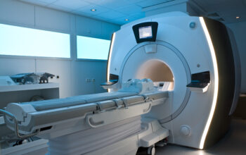
Full-body MRI (Magnetic Resonance Imaging) scans have revolutionized the landscape of medical diagnostics, offering intricate insights into the human body’s anatomy without the need for invasive procedures. This article delineates the operational principles of full-body MRI scans, elucidates the safety considerations associated with their use, and delineates readers’ expectations regarding MRI technology.
At their core, full-body MRI scans harness the principles of nuclear magnetic resonance, exploiting the unique properties of hydrogen nuclei present abundantly in water, which constitutes a significant portion of the human body. When a patient is placed inside the magnetic field of the MRI machine, these hydrogen nuclei align with the magnetic field. This alignment, however, is temporarily disrupted when radiofrequency pulses are applied, causing the hydrogen nuclei to resonate. As the nuclei return to their original alignment, they emit radio signals which are then detected and translated into detailed images of the internal structures.
The process is highly nuanced, encompassing a series of intricate steps. Initially, the patient is positioned within a cylindrical magnet where the magnetic field is most potent. The duration of the scan can vary, typically lasting between 30 to 90 minutes, contingent upon the specificity and comprehensiveness of the imaging required. The machine’s computer processes these radio wave signals to construct high-resolution images, providing a three-dimensional visualization of organs, tissues, and other anatomical structures.
Different sequences can be utilized during an MRI scan, each tailored for specific diagnostic purposes. For instance, T1-weighted images offer superior anatomical detail, allowing for the discernment of fat and water content in tissues, while T2-weighted images excel in depicting fluid distribution, thereby revealing pathology such as tumors or inflammation. Functional MRI (fMRI), a variant of standard MRI, enables clinicians to assess brain activity by measuring cerebral blood flow, enhancing the understanding of neuroanatomical correlations.
The operational nuances of the MRI also allow for the integration of contrasting agents in some examinations. Gadolinium-based contrast agents are administered in specific clinical scenarios to augment the visibility of blood vessels or identify lesions more effectively. However, the use of contrast agents introduces an additional layer of consideration regarding patient safety.
Safety is paramount in medical imaging, and full-body MRI scans are regarded as exceptionally safe modalities due to their non-ionizing nature. Unlike X-rays or CT scans, which expose patients to potentially harmful ionizing radiation, MRI scans utilize magnetic fields and radiofrequency pulses that do not pose the same risk. This intrinsic safety profile makes MRI scans particularly advantageous for imaging in sensitive populations, including pregnant women and pediatric patients.
However, several safety considerations must be addressed. Patients with certain implanted medical devices, such as pacemakers or cochlear implants, may be contraindicated for MRI scans due to the potential for the strong magnetic field to interfere with these devices. It is also crucial for patients to disclose any metallic foreign bodies or a history of metalworking, which could pose risks during the scan.
Rarely, allergic reactions to gadolinium-based contrast agents can occur, necessitating careful screening and monitoring of patients with a history of allergies or kidney dysfunction, as gadolinium may exacerbate renal impairments in susceptible individuals. This necessitates a thorough pre-scan evaluation, wherein clinical history and current health status are meticulously assessed by medical professionals.
With the increasing utilization of MRI technology, various advancements have emerged to enhance efficiency and improve patient comfort. Open MRI machines are specifically designed to alleviate claustrophobic reactions that some individuals may experience in conventional closed MRI systems. These open systems allow for a less constricted scanning space, although the resolution may sometimes differ from that of traditional systems.
Moreover, MRI technology is continuously evolving, and innovations such as artificial intelligence are beginning to augment conventional methods by optimizing image acquisition and interpretation, further stabilizing the role of MRI in diagnostic accuracy. These technological strides enhance the speed and precision of scans, thereby improving outcomes in patient management.
In conclusion, full-body MRI scans serve as a pivotal tool in modern medical diagnostics, providing unparalleled insights into the anatomy and physiology of the human body while prioritizing patient safety. With their non-invasive nature and the absence of ionizing radiation, these scans facilitate the detection of a myriad of conditions ranging from neurological disorders to musculoskeletal injuries. As technology continues to advance, the application of MRI is poised to expand further, promising enhanced diagnostics and personalized patient care strategies.
For those seeking to understand their health or the rationale behind their physician’s referral for an MRI, awareness of the operational principles and safety parameters of this sophisticated imaging technology is indispensable. Patients can approach their medical evaluations with a balanced perspective, informed about the efficacy and safety that full-body MRI scans invariably provide.