Magnetic Resonance Imaging (MRI) scans have revolutionized the field of medical diagnosis, offering unparalleled insights into the human anatomy without the necessity of invasive procedures. This non-invasive imaging technique utilizes powerful magnetic fields and radio waves to generate exquisite images of the internal structures of the body, facilitating accurate diagnosis and treatment planning. As the complexity of the human body continues to astonish the scientific community, the MRI scan stands as a testament to the marvels of contemporary medical technology, making it an object of both practical necessity and deep fascination.
To comprehend the order and precision of an MRI scan, one must first understand the foundational principles upon which this technology is built. At its core, MRI harnesses the inherent properties of hydrogen nuclei—abundant in the human body due to the high water content of tissues. When subjected to a strong magnetic field, these nuclei are aligned to a particular orientation. Upon the application of radiofrequency pulses, they are perturbed from their equilibrium state. As they return to their original alignment, these nuclei emit signals that are captured and converted into detailed images by sophisticated algorithms. This intricate dance of atomic behavior is not merely a scientific curiosity; it encapsulates a profound interconnectedness of physical laws and human health.
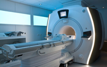
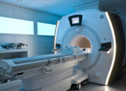
The components of an MRI system play a critical role in the imaging process. The magnet is the heart of the operation, typically a superconducting magnet capable of generating a magnetic field strength of 1.5 to 3.0 teslas—far more potent than that of the Earth’s magnetic field. The gradient coils within the MRI machine enable spatial encoding of the emitted signals, allowing for the precise localization of the tissues being examined. The radiofrequency coils transmit the pulses and receive the returning signals, while the computer system processes this data to produce images that delineate anatomy with stunning resolution. This convergence of engineering and biology underscores the sheer prowess of modern healthcare technologies.
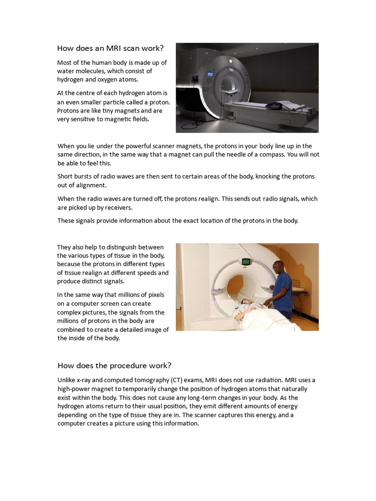
When considering the practicalities of undergoing an MRI scan, patients frequently harbor a range of emotions—from apprehension to intrigue. Typically, the process begins with preparation; patients may be required to change into a hospital gown and remove any metallic items, as these can interfere with the magnetic field. The scan environment, often described as a narrow tunnel, can evoke feelings of claustrophobia. However, it is essential to recognize that advances in design, such as open MRIs, have mitigated this concern for many individuals.
The actual scanning process requires the patient to lie still on a movable table, which is then positioned within the magnet. As the scan commences, intermittent sounds akin to banging or thumping emerge from the machine; these noises result from the rapid switching of magnetic gradients. Patients may be provided with auditory distractions, such as music, to enhance their comfort. Interestingly, while the MRI itself is non-invasive and safe, it is crucial to communicate any pre-existing conditions, such as the presence of pacemakers or certain metal implants, as these can pose significant risks within the magnetic field.
After the scan concludes, radiologists—medical specialists trained in interpreting diagnostic imaging—analyze the acquired images. The adeptness with which they can discern anomalies not visible to the naked eye plays a critical role in diagnosing conditions ranging from tumors and lesions to ligament tears and vascular malformations. The multifaceted imaging capabilities of MRI, including diffusion-weighted imaging and functional MRI (fMRI), enable clinicians to visualize not only static anatomy but also dynamic physiological processes, leading to deeper insights into conditions such as Alzheimer’s disease, stroke, or even psychiatric disorders.
A fascinating aspect of MRI technology lies in its adaptability and the continuous evolution of techniques. Functional MRI, for example, capitalizes on the differences in blood flow associated with neural activity. By measuring changes in cerebral blood flow, fMRI allows researchers to map brain activities during various cognitive tasks, providing a window into the lexicon of human thought and emotion. This blending of neuroscience with advanced imaging heralds exciting possibilities for both research and clinical applications.
As we delve deeper into the implications of MRI technology, we must also consider the ethical dimensions intertwined with its use. Questions regarding access to MRI resources, particularly in under-resourced regions, heighten the ongoing discourse on health equity. The promise of MRI technology must be balanced with the challenge of ensuring that advancements in medical imaging are accessible to all, fostering an inclusive approach to healthcare innovation.
In summary, an MRI scan is not merely a diagnostic tool; it epitomizes the confluence of technology and biology. From the meticulous preparation prior to the imaging to the interpretation of resultant images by highly trained radiologists, each phase of the process underscores a commitment to accuracy and patient care. This intricate blend of science and artistry allows for unparalleled glimpses into the human body, illuminating the pathway towards enhanced understanding and treatment of myriad medical conditions. As MRI technology continues to evolve, so too does the potential for transformative breakthroughs in the realm of health and medicine.