In the realm of medical diagnostics, two modalities reign supreme in their respective applications: Magnetic Resonance Imaging (MRI) and Positron Emission Tomography (PET) scans. Both techniques are pivotal in clinical settings, yet they diverge significantly in their mechanisms, applications, and the types of information they provide. Understanding these differences garners intrigue, as each modality offers unique insights into human anatomy and physiology, probing the depths of the body’s inner workings.
Understanding the Basic Mechanisms
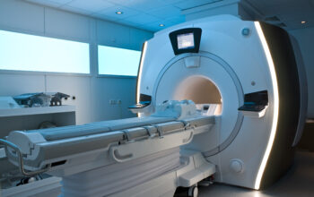
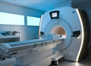
At the core of MRI technology lies the principle of nuclear magnetic resonance. This non-invasive imaging technique exploits the magnetic properties of hydrogen nuclei—predominantly found in water molecules of the human body. When subjected to a strong magnetic field, these protons emit signals that are converted into detailed images by sophisticated software algorithms. This process reveals the structural intricacies of soft tissues, making MRI indispensable in neurology, orthopedics, and oncology.
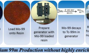
Conversely, PET scans operate on a fundamentally divergent paradigm rooted in nuclear medicine. This imaging modality employs radiopharmaceuticals—tracers that emit positrons—as a means to visualize metabolic processes within the body. Following the intravenous administration of these tracers, a positron is emitted, annihilating with an electron, which subsequently generates gamma rays detectable by the PET scanner. This technique elucidates the functional aspects of cellular activity, thus offering insights into disease detection, particularly in cancer and neurodegenerative disorders.
Key Differences in Imaging Intent
The contrasting intents of MRI and PET scans are foundational to their application in medical diagnostics. MRI is chiefly utilized for anatomical delineation. It excels in visualizing structures such as the brain, spinal cord, joints, and soft tissues. Pathologies such as lesions, herniated discs, and tumors can be effectively characterized through high-resolution images that delineate tissue contrast. The plethora of MRI sequences allows for tailored imaging protocols to suit specific diagnostic inquiries.
In juxtaposition, PET scans are quintessential for assessing physiological and biochemical function within the body. The metabolic information gleaned from PET imaging allows clinicians to characterize tumors by gluco-metabolic activity, assessing malignancy and treatment efficacy. Therefore, while MRI elucidates structure, PET elucidates function, rendering them complementary in comprehensive cancer care management.
Contrast Agents and Safety Profiles
MRI often employs gadolinium-based contrast agents to enhance image contrast, especially in discerning vascular structures and pathological conditions. The safety profile of gadolinium is generally favorable; however, caution is warranted in patients with pre-existing renal insufficiencies, as this could precipitate nephrogenic systemic fibrosis.
Conversely, in the PET realm, the tracers used are radioactive, albeit at low doses. Safety considerations primarily revolve around radiation exposure, a pivotal concern in patient care. The risk of radiation-induced malignancy must be judiciously balanced against the diagnostic utility of the scan. Importantly, advancements in tracer technology and dosage optimization are continually mitigating these risks.
Patient Experience and Procedure Duration
The experience of undergoing MRI versus PET scanning varies considerably. An MRI session typically lasts between 15 to 60 minutes, depending on the complexity of the examination. Patients must remain motionless within a cylindrical magnet, which can incite claustrophobia or anxiety in some individuals. Modern MRI systems have increasingly integrated open designs to alleviate these concerns, though they may compromise spatial resolution.
A PET scan, while generally shorter—averaging around 30 minutes—entails the prior necessity of a preparatory phase wherein the radiotracer is administered. Following injection, a waiting period of approximately 30 to 60 minutes is mandatory to permit the tracer to localize within targeted tissues. This temporal component can add complexity to patient scheduling and overall imaging workflow.
Complementary Roles in Oncology
The intersection of MRI and PET in oncology showcases their synergistic potential. Given their contrasting yet complementary strengths, they are frequently employed in tandem to furnish a comprehensive view of tumor physiology and anatomy. For instance, a patient may receive an MRI to ascertain the structural ramifications of a tumor while simultaneously undergoing PET imaging to evaluate the metabolic aggressiveness of the lesion. The confluence of these modalities can significantly enhance staging accuracy and therapeutic planning.
Future Directions
As medical technology continues to progress, the potential for integrative imaging modalities stands at the forefront of translational research. Emerging techniques such as PET/MRI hybrid scanners aim to fuse the anatomical precision of MRI with the metabolic insight of PET within a single session, promising unparalleled diagnostic capabilities. This convergence of imaging sciences heralds a new era in diagnostic medicine, wherein multimodal imaging could streamline workflows and enhance patient outcomes.
In conclusion, the divergence between MRI and PET scans serves as a testament to the rich tapestry of medical imaging. While both modalities hold significant value in the diagnostic armamentarium, their unique mechanisms and applications cater to varied clinical needs. The fascination surrounding these imaging techniques lies not merely in their technological sophistication but in the profound capacity to illuminate the intricacies of human health—a pursuit that is as captivating as it is crucial in the modern era of medicine.