Understanding MRI: An Overview of Magnetic Resonance Imaging
Magnetic Resonance Imaging (MRI) represents a monumental advancement in medical imaging technology, providing unparalleled insights into the human body’s complex architecture. With its ability to create detailed images of organs, soft tissues, and nerves, MRI plays a pivotal role in diagnostics, treatment planning, and monitoring of various medical conditions. This article delineates the intricate workings of MRI, the different types of MRI scans available, its clinical applications, and the benefits and limitations associated with this sophisticated modality.
1. The Fundamental Principles of MRI
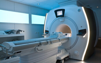
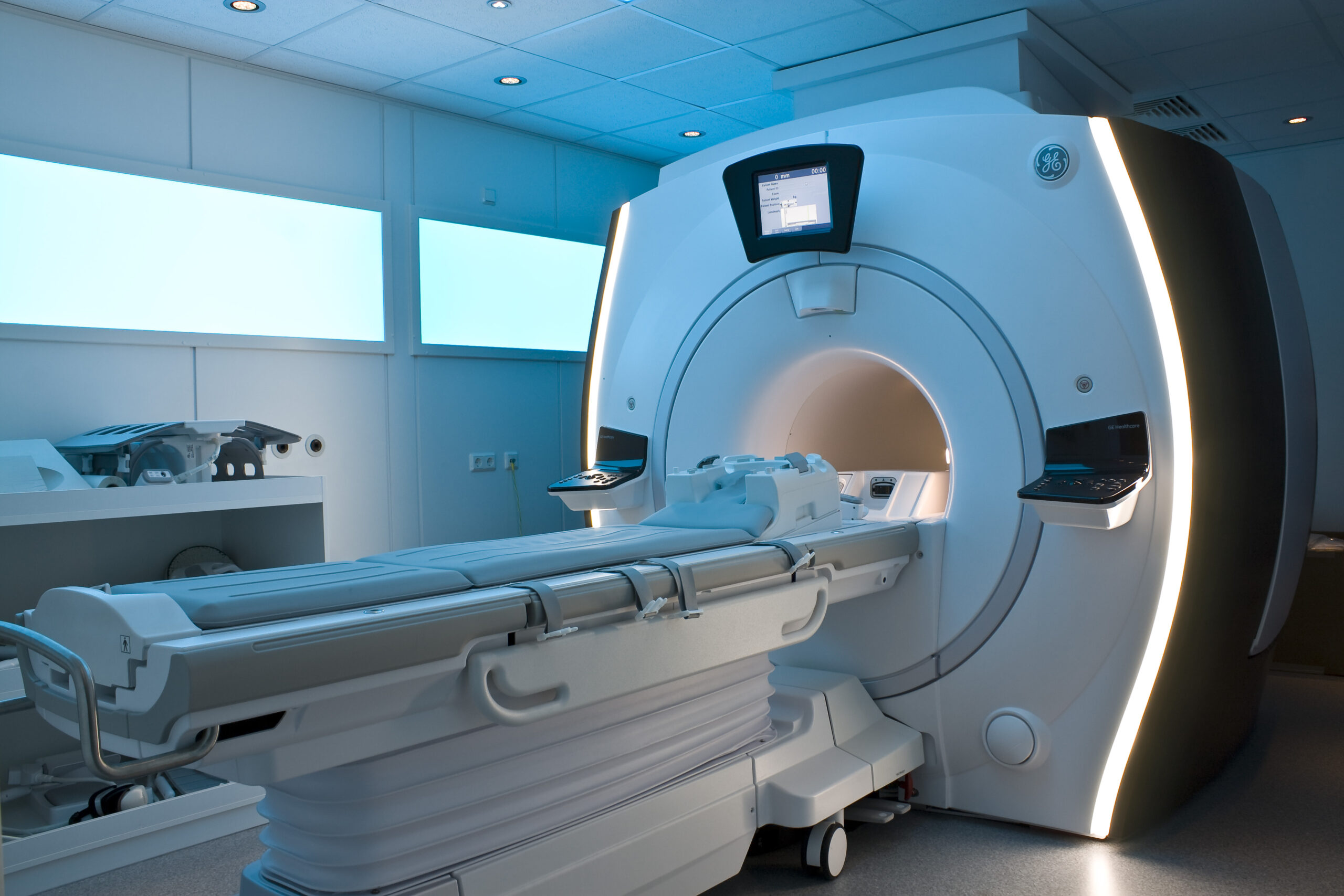
At its core, MRI exploits the magnetic properties of atomic nuclei, particularly those of hydrogen, which is abundantly present in biological tissues due to the high water content in the human body. The technique employs a powerful magnet, which generates a strong magnetic field that aligns hydrogen nuclei. When subjected to radiofrequency (RF) pulses, these aligned nuclei absorb energy and subsequently emit signals as they return to their equilibrium state. This emitted signal is then captured by the MRI scanner and transformed into complex images through sophisticated algorithms.
One of the key advantages of MRI is its ability to manipulate the emitted signals using various techniques, such as T1-weighted and T2-weighted imaging. These contrasts accentuate different properties of the tissues, enabling clinicians to distinguish between healthy and pathological states, thus enhancing the diagnostic accuracy.
2. Types of MRI Scans
There exists an array of MRI techniques tailored to address specific diagnostic needs:
2.1 Standard MRI Scans
The conventional MRI consists of different sequences, mainly T1 and T2 imaging. While T1-weighted images provide insights into anatomical structures with high fat content, T2-weighted images are adept at highlighting fluid-filled areas, rendering them helpful in identifying edema and inflammation.
2.2 Functional MRI (fMRI)
Functional MRI has revolutionized the field by allowing for the visualization of brain activity. It measures fluctuations in blood flow and oxygenation levels, thereby linking specific cerebral activities to particular regions of the brain. This technique is invaluable for neurosurgical planning, revealing eloquent cortex areas that govern crucial functions such as speech and motor control.
2.3 Diffusion Tensor Imaging (DTI)
Diffusion Tensor Imaging is a specialized MRI technique that visualizes white matter tracts in the brain. By measuring the diffusion of water molecules, it provides insights into the integrity and orientation of neural pathways, assisting in the understanding of neurodegenerative diseases and traumatic brain injuries.
2.4 Magnetic Resonance Angiography (MRA)
MRA allows for the non-invasive assessment of blood vessels, utilizing the same principles as MRI to create detailed images of arteries and veins. This technique is particularly beneficial for detecting vascular abnormalities, such as aneurysms or stenoses, without the need for contrast agents, although contrast-enhanced MRA may be employed for enhanced visualization.
3. Clinical Applications of MRI
The clinical applicability of MRI spans numerous medical specialties:
3.1 Neurology
In neurology, MRI is instrumental in identifying various conditions, such as tumors, strokes, multiple sclerosis, and neurodegenerative disorders. Its exquisite detail makes it the gold standard for brain imaging, facilitating accurate diagnosis and management.
3.2 Orthopedics
In orthopedics, MRI is invaluable for assessing soft tissue injuries, joint disorders, and musculoskeletal tumors. Its capability to visualize cartilage, ligaments, and tendons makes it an essential tool in sports medicine and rehabilitation.
3.3 Oncology
In oncology, MRI assists in tumor detection, staging, and treatment monitoring. It provides critical information regarding tumor size, location, and involvement of adjacent structures, aiding in the formulation of treatment plans and evaluation of therapeutic efficacy.
4. Advantages of MRI
The comprehensive benefits of MRI are multifaceted:
- Non-ionizing radiation: Unlike X-rays or CT scans, MRI employs magnetic fields and radio waves, eliminating exposure to ionizing radiation, making it a safer option for repeated imaging.
- High-Resolution Images: MRI produces remarkably high-resolution images with excellent contrast between different types of tissues, rendering it superior for soft tissue evaluation.
- Versatility: With various scan types available, MRI can address a wide range of diagnostic queries, adapting to clinical necessities.
5. Limitations of MRI
Despite its advantages, MRI is not devoid of limitations:
- Cost: MRI procedures can be expensive, making them less accessible for some patients.
- Time-consuming: Scanning times can be lengthy, necessitating patients to remain still, which may be challenging for certain populations.
- Metal Implants: Patients with certain implants, such as pacemakers, may be ineligible for MRI, restricting its utilization in specific cases.
Conclusion
In sum, MRI has fundamentally altered the landscape of medical imaging, allowing for enhanced diagnostic capabilities and better patient outcomes. Its versatility, coupled with the detailed insights it provides, renders MRI an indispensable tool in contemporary medicine. Understanding its mechanisms, applications, and limitations is crucial for both clinicians and patients, ensuring informed decisions regarding patient care and management.