Molecular imaging has emerged as a paramount technique in contemporary biomedical research and clinical practice, allowing researchers and clinicians to visualize biological processes at the cellular and molecular levels. The fascination surrounding molecular imaging stems from its dual ability to provide exquisite detail of biological interactions and to inform therapeutic strategies. This article delineates several prominent modalities of molecular imaging, elucidating their principles, applications, and relevance in both research and clinical settings.
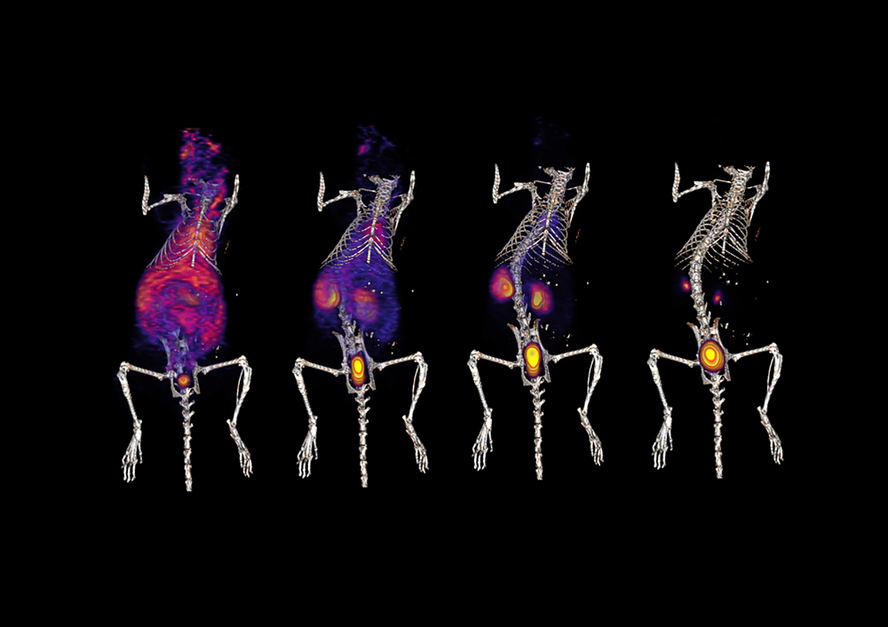
One of the quintessential types of molecular imaging is **Positron Emission Tomography (PET)**. This non-invasive imaging technique utilizes radiotracers—radioactively labeled compounds—that emit positrons upon decay. These emitted positrons interact with electrons, leading to the production of gamma rays that are detected by a PET scanner. The ability to visualize metabolic processes in real time has rendered PET indispensable in oncology, neurology, and cardiology. For instance, PET is particularly effective in identifying malignant tumors, as cancerous cells often exhibit heightened glucose metabolism, easily detectable due to the radiolabeled glucose analog, fluorodeoxyglucose (FDG). This capability transforms not only diagnostics but also therapeutic monitoring, offering insights into treatment efficacy.
Another pivotal modality is **Single Photon Emission Computed Tomography (SPECT)**. SPECT operates on a principle similar to that of PET but utilizes gamma cameras to capture emissions from radiolabeled compounds that have been introduced into the body. While SPECT may not provide the same level of spatial resolution as PET, it excels in functional visualization and is particularly beneficial for cardiac imaging. Agents such as technetium-99m are employed to assess blood flow and perfusion in myocardial tissues, facilitating the diagnosis of coronary artery disease and myocardial infarction. The relatively lower cost and greater availability of SPECT make it a practical choice in various clinical settings.
**Magnetic Resonance Imaging (MRI)**, while traditionally considered a structural imaging modality, has evolved to encompass molecular imaging through the implementation of specific contrast agents. The advent of advanced techniques such as Magnetic Resonance Spectroscopy allows for the evaluation of metabolic processes within tissues. For example, the use of hyperpolarized agents can illuminate metabolic pathways in vivo, thereby improving our understanding of diseases like cancer and neurodegenerative disorders. The exquisite soft tissue contrast afforded by MRI, combined with its high spatial resolution, underscores its significance in non-invasive diagnostics.
Furthermore, **Optical Imaging** has garnered considerable attention due to its remarkable sensitivity and versatility. Techniques such as bioluminescence and fluorescence imaging leverage light emission from either genetically engineered proteins or injected fluorescent dyes to visualize molecular events. Optical imaging is particularly advantageous in small animal models, where its ability to provide real-time imaging of biological processes enhances the understanding of disease progression and treatment effects. However, its predominantly superficial penetration limits applications in large animals and humans, necessitating innovative approaches to overcome these challenges.
In addition to these established modalities, **Theranostics**, a term combining therapy and diagnostics, has emerged as a cutting-edge approach where molecular imaging directly informs therapeutic decisions. This paradigm utilizes targeted radiopharmaceuticals that are designed for both imaging and treatment. A salient example of this is seen in prostate cancer, where agents like ^223Radium can be employed to visualize and treat metastatic lesions simultaneously. The integration of imaging with therapeutic interventions marks a significant advance in personalized medicine, aligning treatment strategies more closely with individual patient profiles.
Moreover, **Computed Tomography (CT)**, although chiefly recognized for its structural imaging capabilities, is oftentimes utilized in conjunction with molecular imaging techniques. For example, hybrid imaging systems such as PET/CT amalgamate the functional insights of PET with the anatomical details from CT, enhancing diagnostic accuracy. Such synergistic approaches enable clinicians to not only pinpoint the location of a metabolic abnormality but also to discern its anatomical context, which is critical for guiding interventions.
The evolution of molecular imaging techniques continues to be driven by technological innovations. **Nanoimaging**, which employs nanoparticles as contrast agents, is one of the most exciting frontiers in this domain. Nanoparticles can be engineered to possess unique optical and magnetic properties, granting unprecedented sensitivity and specificity to molecular targets. This paradigm shift is poised to enhance the early detection of diseases, particularly cancer, by enabling the identification of pathological changes well before clinical symptoms manifest. The potential for nanoparticles to deliver therapeutic agents in a targeted manner further amplifies their clinical relevance.
In summation, molecular imaging encompasses a diverse array of modalities that collectively revolutionize our capacity to interrogate biological systems at the finest scales. The integration of these techniques into clinical practice offers profound implications for diagnosis, treatment, and ultimately, patient outcomes. As advancements in imaging technology continue to unfold, the anticipation surrounding molecular imaging burgeons, ensuring its critical role in the future landscape of medicine. The profound implications of these methodologies extend beyond mere visualization, guiding researchers and clinicians toward a more nuanced understanding of complex biological phenomena.