The advent of computed tomography (CT) scans revolutionized diagnostic medicine, providing unprecedented images of internal anatomy. However, their widespread utility brings a critical question to the forefront: how does the radiation exposure inherent in CT scans impact health, particularly with repeated examinations? As we delve into the implications of this “silent dose,” one must ponder: have we become desensitized to the risks of imaging technologies?
While the benefits of CT scans in diagnosing conditions are irrefutable, a nuanced understanding of the associated radiation risks is essential. Unlike conventional X-rays, CT scans utilize a series of rotating X-ray images to create detailed cross-sectional views, which often results in a significantly higher radiation dose. This higher dose can become a cause for concern, especially when juxtaposed with long-term health outcomes.
**1. Understanding Radiation Exposure**
Each CT scan delivers a radiation dose measured in millisieverts (mSv). For context, a single CT scan of the abdomen can administer up to 10 mSv, whereas a standard chest X-ray is merely around 0.1 mSv. The cumulative effects of exposure become more pronounced with increased frequency of scans, leading to queries about a patient’s radiation history and potential dose accumulation.
**2. The Risk Paradigm: The Linear No-Threshold Model**
The prevailing paradigm for evaluating radiation risk is the Linear No-Threshold (LNT) model. This model postulates that even the smallest doses of ionizing radiation can incrementally increase the risk of cancer. When patients consider their individual risk factors—age, gender, pre-existing conditions—the hunt for an optimal balance between diagnostic benefit and risk becomes a rigorous exercise in prudence.
**3. Who is Most at Risk?**
Certain populations warrant heightened vigilance. Children, for instance, are more susceptible to radiation-induced malignancies due to their developing tissues and longer life expectancy. Likewise, individuals with pre-existing conditions necessitating frequent imaging may encounter escalated risk profiles. In these cases, one must meticulously assess the necessity and frequency of CT scans, probing where alternatives might be employed without sacrificing diagnostic accuracy.
**4. Alternatives to CT Scans: Weighing the Possibilities**
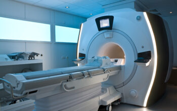
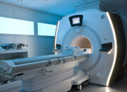
The medical community is increasingly advocating for non-ionizing imaging modalities, such as ultrasound and magnetic resonance imaging (MRI). These alternatives provide significant advantages, including the absence of ionizing radiation. However, they may also come with limitations such as longer acquisition times, less precision in some contexts, or limited availability based on geographic and financial factors. Clinicians must engage in thoughtful discussions with patients, guiding them through the risks and benefits of each modality.
**5. Patient Empowerment and Informed Consent**
In this landscape of diagnostic ambiguity, patient empowerment emerges as a critical component. Individuals should feel adequately informed about the necessity of any imaging study before consenting to it. Engaging in discussions that examine prior medical history and potential risks is essential. Patients should feel emboldened to ask questions, such as: “Is this scan absolutely necessary?” or “Are there safer alternatives available?” This dialogue fosters a partnership between healthcare providers and patients, ensuring that decisions are made collaboratively.
**6. The Role of Technology and Innovation**
Innovations in imaging technology hold promise for reducing radiation doses without compromising diagnostic efficacy. Techniques such as iterative reconstruction algorithms and advanced detectors are being developed to refine image quality while minimizing exposure. As these technologies evolve and become more widely integrated into clinical practice, patients and practitioners alike can hope for a future where the ‘silent dose’ of radiation becomes less of a concern.
**7. Regulatory Oversight and Best Practices**
The regulation of radiation doses in medical imaging is an ongoing concern among health organizations. Guidelines established by various health authorities advocate for the As Low As Reasonably Achievable (ALARA) principle, emphasizing that providers strive to minimize radiation exposure wherever possible. Implementing standard operating procedures within medical facilities further ensures that patients receive optimal care, balancing diagnostic needs with safety.
**8. Moving Forward: Fostering Awareness and Education**
Raising awareness about radiation dosage and reinforcing educational initiatives within the healthcare community is paramount. Continuing medical education programs should include discussions on the implications of radiation exposure, fostering a culture of safety among practitioners who influence when and how imaging modalities are utilized. Such educational efforts can serve to illuminate the conversation surrounding the potential future landscape of imaging diagnostics.
In conclusion, as we consider the implications of the “silent dose” associated with CT scans, stakeholders must remain vigilant. The interplay between diagnostic necessity and potential risk requires careful contemplation, informed consent, and a commitment to progressive methodologies. By enhancing dialogue, prioritizing patient education, and embracing innovation in technology, the medical community can navigate the complex terrain of imaging safety, ultimately ensuring patient welfare remains at the forefront of diagnostic advancement.