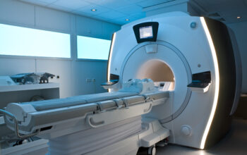
Magnetic Resonance Imaging (MRI) has revolutionized the domain of medical diagnostics, particularly in the realm of neuroimaging. Often, the terms “brain scan” and “MRI” are used interchangeably in colloquial conversations. However, this semantic ambiguity warrants a deeper exploration into both concepts to delineate their distinctions and interrelations. Comprehending these differences not only enhances our understanding of neuroimaging techniques but also illuminates the complexities of brain anatomy and pathology.
To commence, we must define the terminologies clearly. An MRI refers to a specific imaging technique that utilizes strong magnetic fields and radio waves to produce detailed images of the body’s internal structures, particularly useful for visualizing soft tissues such as the brain. Conversely, a brain scan is a broader term that could encompass various techniques employed to visualize and assess the brain’s structure and function. Consequently, while all MRIs can be considered brain scans when applied to the brain, not all brain scans are MRIs. This nuance is fundamental in understanding the landscape of neurological imaging.
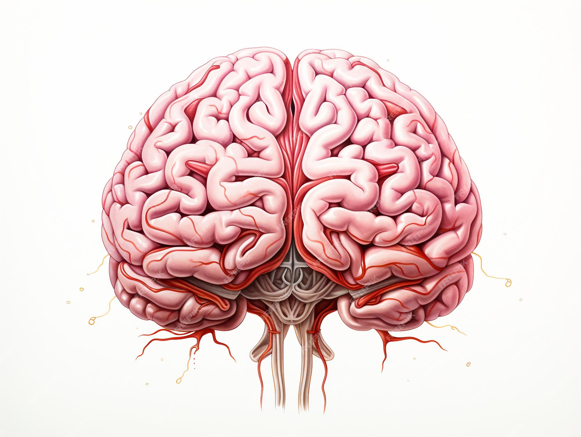
MRI encompasses several subclasses, including functional MRI (fMRI) and diffusion tensor imaging (DTI). Traditional MRI primarily focuses on the anatomical structure of the brain, revealing details of the neural architecture. In contrast, fMRI gauges cerebral blood flow, thus providing insights into brain activity by highlighting regions that demonstrate increased oxygen consumption. This distinction underscores a pivotal dimension of neuroimaging; the relationship between structure and function is complex and multifaceted. By employing various MRI techniques, clinicians and researchers can parse out these intricacies effectively.
Furthermore, brain scans extend beyond MRI into other methodologies, such as computed tomography (CT) scans and positron emission tomography (PET) scans. CT scans employ X-ray technology to generate cross-sectional images of the brain, making them advantageous in acute settings where rapid diagnosis is critical, such as detecting hemorrhagic strokes. PET scans, on the other hand, utilize radioactively labeled tracers to visualize metabolic processes within the brain, offering a unique perspective on neurological diseases such as Alzheimer’s and other dementias.
As we navigate these various modalities, it becomes evident that each imaging technique possesses distinct attributes tailored to specific diagnostic inquiries. The choice between these techniques is often dictated by the clinical context, patient history, and the specific information sought by the healthcare provider. An MRI might be favored for chronic neurological conditions due to its superior soft tissue contrast, whereas a CT scan might be prioritized in emergency scenarios where swift decision-making is paramount.
Understanding the physiological principles underpinning MRI technology can further elucidate why it is often the gold standard for neuroimaging. MRI operates on the principles of nuclear magnetic resonance, which exploits the magnetic properties of atomic nuclei. When subjected to a strong magnetic field, certain nuclei resonate, and with the application of radiofrequency pulses, energy is emitted that can be captured to create detailed images. This non-invasive technique does not utilize ionizing radiation, a feature that appeals to both patients and clinicians, particularly in scenarios involving repeated imaging.
Moreover, the ramifications of MRI extend far beyond mere diagnostics. Research has demonstrated how MRI technologies can catalyze advances in neurological understanding, as they permit non-invasive explorations of the human brain in both health and disease. Studies utilizing fMRI have illuminated the neural correlates of cognition, emotion, and behavior, bridging the domains of neuroscience and psychology. The fascination surrounding MRI technology lies in its ability to render the invisible intricacies of the human mind visible, opening avenues for both scientific inquiry and therapeutic intervention.
Nonetheless, it is imperative to acknowledge the limitations and challenges associated with MRI and other brain imaging techniques. High costs, accessibility issues, and the necessity for patient compliance can impede widespread adoption. Additionally, while MRI is exceptionally adept at revealing structural anomalies, its utility in identifying certain pathologies, such as acute ischemic strokes, can be limited. Therefore, an integrative approach that synthesizes information across various imaging modalities often yields the most comprehensive understanding of complex neurological conditions.
Furthermore, emerging advancements in neuroimaging are propelling the field into unprecedented territories. Innovations such as machine learning algorithms for image analysis present the possibility of enhancing diagnostic accuracy. These technologies can parse vast datasets, identifying subtle patterns that might elude the human eye. The intersection of technology and medical imaging heralds a new epoch in neuroscience, where early detection and tailored therapies may proliferate.
In conclusion, while the term “brain scan” broadly encompasses various imaging modalities, MRI is a specific technique that stands out due to its remarkable anatomical detail and functional capabilities. Understanding the nuances between these terms enriches our appreciation of the complexities inherent in brain imaging. As neuroscience continues to evolve, the fascination surrounding imaging techniques is likely to burgeon, as they serve not only as diagnostic tools but as windows into the enigmatic workings of the human brain.