Magnetic Resonance Imaging (MRI) has revolutionized the field of medical diagnostics, serving as a crucial tool for visualizing internal bodily structures without the necessity of invasive procedures. Operating an MRI machine is a sophisticated process that requires not only technical acumen but also an understanding of the underlying physics principles that govern its operation. This article will delineate the multifaceted approach to operating an MRI machine, addressing critical operational procedures, safety protocols, and the intricate interplay of technology and patient care.
Understanding the MRI Machine Components
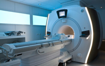
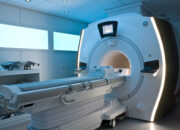
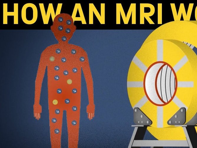
Before delving into the operational intricacies, it is paramount to comprehend the primary components of an MRI machine. Key elements include the magnet, radiofrequency coils, gradient coils, and the computer systems that process the obtained data. The magnet, typically a superconducting electromagnet, creates a strong and uniform magnetic field that is essential for aligning hydrogen nuclei (protons) in the body. Radiofrequency coils are crucial for transmitting and receiving radio waves, while gradient coils enable spatial localization of the MRI signals.
The magnet stands as the core of the MRI machine. Its strength is expressed in teslas, with clinical machines generally ranging from 1.5 to 3.0 teslas. The magnetic field generated is critical for the resonance phenomenon that underpins MRI imaging. The interplay of these various components is fundamental to acquiring high-quality images.
Preparing the Patient
Effective operation of an MRI machine extends beyond technical expertise; it requires a comprehensive understanding of pre-examination protocols aimed at ensuring patient safety and comfort. Prior to scanning, the patient must undergo a thorough screening process to identify contraindications such as the presence of ferromagnetic implants or devices, which can lead to severe complications in the magnetic field of the MRI scanner.
Once cleared, the next step involves educating the patient about the procedure, incorporating a detailed explanation of the scanning process, duration, and potential sensations, such as loud noises emitted by the gradient coils. This preparatory phase is vital in alleviating any anxiety the patient may have. Comfortable positioning is facilitated by the use of specialized cushions and supports, ensuring that the patient remains still during the imaging process to enhance the image quality.
Operational Protocols for MRI Scanning
Initiating the MRI procedure necessitates meticulous adherence to operational protocols, which can be categorized into several key steps:
- Calibration of the MRI System: Calibration involves setting up the machine according to manufacturer specifications, ensuring that the magnetic field and gradient coils are functioning optimally. This is often performed by the technologist before any patient is scanned.
- Patient Positioning: The patient is carefully positioned on the table, with the specific area of interest aligned at the center of the magnet bore. Securely strapping the patient may be necessary to prevent movement during imaging.
- Setting Imaging Parameters: The technologist must meticulously configure imaging parameters such as repetition time (TR), echo time (TE), and slice thickness. This requires a solid understanding of how these values influence image clarity, contrast, and resolution.
- Monitoring During the Scan: As the scan is conducted, constant monitoring of the patient’s condition is essential. Communication is maintained through intercom systems, allowing the technologist to ensure the patient’s comfort and address any concerns promptly.
Data Acquisition and Processing
The data acquisition phase is where the complexity of MRI technology manifests itself. Upon interaction with the magnetic field and radiofrequency pulses, protons emit signals that are contingent upon their local environment, which is based on tissue type, water content, and pathological states. The resulting data is then digitized and subjected to Fourier transformation, a mathematical process that translates the raw data into usable images.
Post-processing techniques allow for further refinement of the images. Advanced algorithms can enhance image contrast, reduce noise, and apply three-dimensional reconstruction, providing clinicians with a more holistic view of the anatomy under examination. The resulting images can also be modified to highlight various tissues, such as fat suppression sequences that isolate lesions from surrounding adipose tissue.
Safety Protocols and Magnetic Field Considerations
Safety management is a cornerstone of MRI operation. The strong magnetic fields can attract ferromagnetic objects with significant force, necessitating the implementation of strict protocols. Patients and personnel must be screened for metallic objects, and “zone” management is enforced to delineate areas of varying magnetic risk. Additionally, magnets pose risks related to projectiles and heating from radiofrequency exposure, necessitating adherence to safety guidelines.
Furthermore, the role of the imaging team extends to ensuring compliance with established safety standards while effectively managing any potential emergencies, such as patient distress or an adverse reaction to contrast agents. Emergency protocols must be well-understood by all staff members involved in patient care during an MRI scan.
Conclusion
Operating an MRI machine is not merely a matter of technical prowess but intertwines sophisticated engineering, physics principles, patient engagement, and rigorous safety protocols. The interplay of these elements highlights the broader significance of MRI technology in contemporary medicine, providing invaluable diagnostic insights that facilitate timely and accurate patient care. Furthermore, as technological advancements continue to unfold, the potential for MRI applications to expand into new realms of clinical practice is immense, promising an exciting future at the intersection of technology and healthcare.