Radiology presents a fascinating intersection of science and art, a realm where the invisible becomes visible through the interplay of physics and technology. As medical imaging burgeons into an essential tool for diagnosis and treatment, an understanding of the underlying physics in radiology is not only beneficial but imperative for those aspiring to master this field. However, how much physics is truly required for a radiology course? The answer can be likened to peeling layers of an onion; each layer reveals complexity, nuance, and a profound connection to the discipline itself.
The foundations of radiologic physics are built upon principles that govern energy and matter. At the core lies a fundamental grasp of atoms and molecules, which serve as the building blocks of all substances, including biologic tissues. A radiologist must comprehend how these minute entities interact with various forms of radiation, such as X-rays, to produce images that convey critical diagnostic information. The atomic structure, characterized by electrons, protons, and neutrons, is essential, as the behavior of these particles under radiation influences image quality and patient safety.
To embark on a comprehensive study of radiologic physics, one must first familiarize themselves with the nature of electromagnetic radiation. This spectral orchestra encompasses a multitude of wavelengths, ranging from radio waves to gamma rays. In the context of radiology, the X-ray portion of the spectrum plays a pivotal role. It is imperative to understand how photons, the quantized units of electromagnetic radiation, are produced and manipulated. Specific emphasis is often placed on X-Ray production, wherein electrons are accelerated and directed to a metal target, resulting in the emission of high-energy photons. This phenomenon is not merely a technicality but a foundation upon which radiographic imaging rests.
Moreover, an exploration of the interactions between radiation and matter becomes crucial. When X-rays traverse through human tissue, they encounter varying degrees of attenuation based on tissue density and composition. This interaction is explored through concepts such as the photoelectric effect and Compton scattering, which elucidate how different tissues absorb or scatter photons. An adept radiologist must interpret these interactions to differentiate between normal and pathological states within the body. Thus, a firm grounding in these principles equips future radiologists with the analytical skills necessary to understand the nuances of diagnostic imaging.
In addition to understanding electromagnetic radiation, the principles of wave-particle duality unveil the complexities of X-ray imaging. This concept articulates that radiation possesses both wave-like and particle-like properties, leading to phenomena such as interference and diffraction, which can affect image clarity. The mathematical manifestations of these principles, often conveyed through wave functions and Fourier transforms, become vital tools in the radiologist’s toolkit when optimizing imaging techniques to enhance diagnostic efficacy.
Furthermore, one must delve into the aspects of image formation and processing, which are paramount in today’s radiologic practices. Digital radiography, for instance, employs intricate algorithms that convert raw data into interpretable images. Understanding the physics behind digital signal processing, including sampling theory and quantization, is essential for deciphering how information is represented and enhanced in a way that aids clinical diagnoses. Each pixel on a digital image embodies a repository of information that reveals the delicate interplay between dose and image quality, a crucial consideration in ensuring patient safety while maximizing diagnostic yield.
Another integral component of radiologic physics is the calculation of dose and radiation safety. The principle of ALARA—”As Low As Reasonably Achievable”—serves as a guiding mantra for radiologists, ensuring that the exposure to ionizing radiation is minimized without compromising diagnostic quality. A thorough understanding of dosimetry, which encompasses concepts such as absorbed dose, effective dose, and dose equivalence, equips future practitioners with the knowledge necessary to assess and mitigate risks associated with various imaging modalities. This mastery not only protects patients but also safeguards healthcare providers in their professional practice.
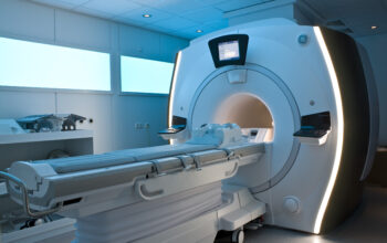
Lastly, as medical technology advances, so too does the significance of emerging imaging modalities, such as computed tomography (CT) and magnetic resonance imaging (MRI). Each of these techniques intertwines physics with innovation, incorporating unique principles to achieve imaging that transcends traditional boundaries. CT relies on the principles of X-ray attenuation and cross-sectional imaging, while MRI employs strong magnetic fields and radio waves, showcasing the diverse applications of physical principles in imaging. Gaining insight into the physics that underlie these technologies enables radiologists to stay abreast of advancements and implement them effectively in patient care.
In conclusion, embarking on a journey through radiologic physics offers an intellectually stimulating experience that intertwines the theoretical with the practical. From the microscopic interactions of particles to the macroscopic interpretations of images, understanding physics is an indispensable pillar of radiology. It empowers medical professionals to harness advanced technologies, ensures radiographic safety, and ultimately enhances patient outcomes. Thus, the physical principles underlying radiology illuminate the path forward, guiding practitioners as they navigate the ever-evolving landscape of medical imaging.