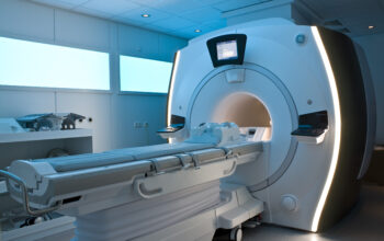
Magnetic Resonance Imaging (MRI) stands as a cornerstone in modern medical diagnostics, enveloping a realm of intricate scientific principles and innovative technology. It transcends traditional imaging modalities by harnessing magnetic fields and radio waves to produce detailed images of the internal structures of the body. This technique invites a deeper contemplation of the human anatomy, unraveling mysteries that conventional imaging could scarcely penetrate.
At its core, MRI employs a potent magnet, which generates a strong magnetic field, typically ranging from 1.5 to 3.0 tesla. This field is significant, as it aligns the protons within the body’s hydrogen atoms, predominantly found within water and fat molecules. When exposed to the magnetic field, these protons resonate at a specific frequency. Subsequently, a pulse of radiofrequency energy is administered, displacing the protons from their alignments. Upon cessation of this pulse, the protons revert to their original state, emitting signals that are subsequently captured and processed by sophisticated computer algorithms to render high-resolution, cross-sectional images of the body.
The fascination with MRI extends beyond its technical foundation; it resides in the myriad of applications it affords across various medical specialties. For instance, in neurology, MRI is indispensable for diagnosing conditions such as multiple sclerosis, brain tumors, and cerebrovascular diseases. The ability to visualize subtle differences in the brain’s morphology and pathology offers unparalleled insights into the complexity of neurological disorders.
Moreover, in orthopedic medicine, MRI emerges as a vital diagnostic tool for evaluating joint and soft tissue injuries. By facilitating detailed imaging of ligaments, muscles, and cartilage, it enables practitioners to ascertain the severity of injuries and craft appropriate treatment plans. This capacity to visualize conditions previously obscured by traditional radiography has revolutionized patient care within this domain.
In the context of oncology, MRI plays a pivotal role in tumor characterization and staging. Enhanced by specific contrast agents that can delineate vascularity and perfusion, MRI can provide nuanced information about tumor size, shape, and aggressiveness. This information is crucial for oncologists in determining not only the treatment modalities but also the prognostic outlook for patients.
One striking attribute of MRI technology is its ability to visualize functional processes within the body, an examination known as functional MRI (fMRI). This variant of MRI exploits the changes in blood flow and oxygenation levels associated with neural activity, thus offering insights into brain function in real-time. fMRI has catalyzed advancements in cognitive neuroscience, further elucidating our understanding of mental processes, cognitive functions, and the underlying architecture of human thought and behavior.
Another compelling facet of MRI is its non-invasive nature, providing a safe alternative to ionizing radiation utilized in computed tomography (CT) scans and X-rays. This aspect is particularly advantageous for vulnerable populations, including pediatric patients and pregnant women, who may be at heightened risk from radiation exposure. The gentleness of the MRI process beckons an era of imaging that prioritizes patient safety while yielding extensive and detailed data.
However, the employment of MRI is not without its challenges and considerations. The strong magnetic fields necessitate stringent safety protocols; individuals with pacemakers, metallic implants, or other ferromagnetic materials may face prohibitive risks. Additionally, the claustrophobic nature of the MRI machine may provoke anxiety in certain patients. Technological advancements, including open MRI systems, aim to mitigate these concerns, though they may not yet fully match the imaging quality of traditional closed systems.
As researchers continue to innovate, the future of MRI holds tremendous promise. Enhanced imaging techniques, such as diffusion tensor imaging (DTI), allow for the examination of white matter tracts in the brain, contributing valuable insights into neurological diseases and injuries. Furthermore, the integration of artificial intelligence in MRI analysis offers the potential for more accurate interpretations, thereby enabling radiologists to make more informed decisions.
In light of these advancements, a curiosity emerges regarding the implications of MRI technology on the broader landscape of healthcare. MRI not only serves the immediate purpose of diagnosis but also catalyzes interdisciplinary collaboration between radiologists, neurologists, surgeons, and oncologists, fostering a holistic approach to patient management. The ability to obtain comprehensive data empowers healthcare professionals, leading to improved treatment outcomes and enhanced patient-centered care.
In conclusion, MRI is far more than a mere diagnostic tool; it symbolizes a shift in our understanding of human anatomy and pathology. By engaging with this sophisticated technology, one transcends the surface-level knowledge of the human body’s structure, delving into the intricate relationships between various systems. MRI hones our curiosity about the unseen, the hidden pathways of disease, and the resilience of the human form. As we stand on the precipice of this technological revolution in medicine, the journey of discovery continues, spurring a relentless quest to explore and understand the intricacies of life itself.


