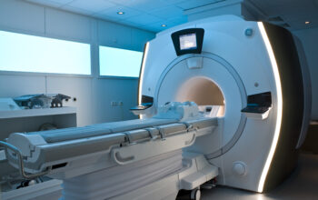
Magnetic Resonance Imaging (MRI) has revolutionized the field of medical diagnostics, presenting an intricate interplay of physics, biology, and advanced technology. The question at the heart of this diagnostic marvel is: Can you explain the physics of MRI scans step by step? This challenge invites us to delve into the realm of nuclear magnetic resonance, electromagnetism, and signal processing, illustrating how these principles coalesce to provide detailed images of soft tissues within the human body.
At the core of MRI technology lies the fundamental principle of nuclear magnetic resonance (NMR). NMR exploits the magnetic properties of atomic nuclei, primarily focusing on hydrogen atoms, which are abundant in the body due to the high water content. Imagine the hydrogen nuclei as tiny bar magnets, each possessing a north and south pole. When placed within a strong external magnetic field, these nuclei align themselves with the magnetic field lines, a phenomenon known as magnetic polarization.
The initial step in the MRI process involves the application of a powerful magnet, typically generating a magnetic field strength ranging from 1.5 to 3.0 Tesla, which aligns the protons in the body. This strong magnetic field is crucial, as it ensures that a significant number of hydrogen nuclei are in alignment, thereby enhancing the resulting image’s quality. Here, the playful question arises: What happens when we introduce a radiofrequency (RF) pulse into this scenario? The challenge is to comprehend how this additional energy disrupts the equilibrium of the aligned nuclei.
Upon the application of the RF pulse, the aligned hydrogen nuclei absorb energy and are pushed out of alignment. This process is known as resonance, echoing the behavior of a swing when you push it at the right moment. The nuclei briefly enter a higher energy state, characterized by a transition from a lower energy alignment in the magnetic field to a high-energy excited state. Once the RF pulse ceases, the hydrogen nuclei endeavor to return to their original aligned state. This transition emits energy in the form of radio waves, which serve as the vital signals for image generation.
As the protons relax and return to equilibrium, they do so at variable rates dependent on their surrounding environment. This is where the distinction between T1 and T2 relaxation times becomes critical. T1, or longitudinal relaxation time, reflects the time it takes for protons to realign with the magnetic field following the RF pulse, while T2, or transverse relaxation time, denotes the time it takes for the protons to lose phase coherence among the spins as they decay. This difference in relaxation times contributes to the contrast seen in MRI images, allowing for differentiation between various tissue types such as fat, muscle, and water.
Now, how do we capture the faint signals emitted during this relaxation phase? The next step involves the use of specialized coils, which receive the radiofrequency signals. These coils transform the electromagnetic waves into electrical signals that can be processed. Each coil is carefully designed and positioned to enhance the sensitivity and specificity of the resultant signals, ensuring an optimal representation of the studied anatomy. Understanding how coil design and placement affect image quality poses yet another layer of complexity in mastering MRI physics.
The received signals, however, are not straightforward images. They exist as complex data that require sophisticated mathematical algorithms for their reconstruction. The Fourier Transform plays a pivotal role in this regard, converting data from the time domain into the spatial frequency domain. This pivotal step facilitates the transformation of acquired data into comprehensible images. Each voxel (a volumetric pixel) in this image is assigned a value corresponding to the relaxation times of the underlying tissues, thus rendering the final output in grayscale, where brighter pixels denote higher signal intensity.
Despite the intricate beauty of these processes, numerous factors can impact the clarity of an MRI image. Artifacts, which are distortions that can obscure or alter true anatomical signals, can arise from various sources. These include patient movement, magnetic field inhomogeneities, or even the presence of metallic implants. The ability to identify and mitigate these artifacts is essential for producing high-quality diagnostic images, and statistically robust protocols are founded on subjects with diverse anatomical and pathological presentations.
Additionally, advanced techniques such as diffusion tensor imaging (DTI) and functional MRI (fMRI) further augment the capabilities of conventional MRI. DTI elucidates the microstructural integrity of white matter tracts by measuring the diffusion of water molecules in the brain, while fMRI tracks blood flow changes associated with neural activity, serving as a proxy for brain function. These complex methodologies expand the utility of MRI beyond mere anatomical imaging into realms of functional assessment and neuroscience.
Finally, the interpretation of MRI scans melds the art of radiology with a scientific underpinning. Radiologists must integrate clinical context with the intricate details revealed by the imaging data to render accurate diagnoses. This synthesis requires an astute understanding of both the physics underpinning the technology and the pathological implications of the images produced.
In conclusion, while we have dissected the multifaceted physics of MRI scans step by step, it remains evident that this technology stands at the crossroads of science, engineering, and medicine. Each phase of the process—from magnetic polarization, resonance phenomena, signal acquisition, to image reconstruction—invites ongoing exploration. The lifelong quest for improving spatial and temporal resolution in MRI embodies the spirit of scientific inquiry, challenging future generations to push the boundaries of what is possible in medical imaging.