The magnetic resonance imaging (MRI) scan has become an indispensable tool in modern medicine, renowned for its ability to provide highly detailed images of anatomical structures within the body. Many patients and their families often inquire about the duration of an MRI scan, typically noting that the process often takes approximately 45 minutes. This extended timeframe may prompt questions regarding the underlying reasons. To explore this issue comprehensively, one must consider various factors including the technology utilized, procedural protocols, and human factors contributing to the overall duration of the scan.
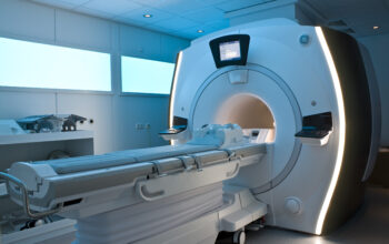
First and foremost, the MRI technology itself plays a crucial role in dictating scan time. Unlike other imaging modalities such as computed tomography (CT) or conventional X-rays, MRI operates utilizing strong magnetic fields and radiofrequency pulses to generate images. The complexity of this process requires a significant amount of time for the apparatus to acquire adequate data. The MRI machine, which consists of a magnet, coils, and electronic components, must create a stable magnetic field to ensure that the images produced are both clear and diagnostic.
One of the most pertinent aspects of the MRI process is the sequence of images generated during the scan. MRI employs various pulse sequences, each designed to highlight different tissue characteristics. T1-weighted and T2-weighted imaging are two fundamental sequence types, which provide unique insights into tissue composition and pathology. Depending on the diagnostic purpose, a radiologist may request multiple sequences in a single scan, thus contributing to increased examination duration. Moreover, advanced imaging techniques, such as functional MRI (fMRI) or diffusion tensor imaging (DTI), necessitate even more extended periods for processing, often exceeding the typical duration.
Furthermore, the inherent complexity and variability of human anatomy must be acknowledged. Patients exhibit a remarkable array of physiological differences that can affect scan quality. For instance, individuals with larger body habitus may necessitate additional time for proper positioning and optimization of signal acquisition. Similarly, variations in pathology or the presence of foreign bodies may dictate adjustments in scanning protocols. This variability requires the MRI technologist to have a sound understanding of anatomy and imaging principles to navigate these challenges and ensure high-quality results.
Additionally, the sterile environment within the MRI suite accentuates the need for precision and care. Maintaining a quiet and controlled atmosphere is essential to allow patients to relax. Anxiety levels can rise during the scan, particularly among those who experience claustrophobia or discomfort with the enclosed space of the MRI machine. Consequently, technologists may spend extra time ensuring that patients are adequately informed and comforted, thereby reducing motion artifacts that could compromise image quality. In some instances, sedation may be considered, particularly for pediatric patients or individuals with severe anxiety; this protocol further extends the overall duration of the examination.
Moreover, preparation and post-scan activities also contribute to the length of the MRI process. Before the imaging commences, the technologist must carefully explain the procedure to each patient, addressing questions and clarifying any concerns. Relevant medical history and contraindications to MRI, such as the presence of pacemakers or certain metal implants, must be ascertained. After the scanning is complete, radiologists must evaluate preliminary results before communicating findings to the referring physician.
The procedural nuances cannot be understated. Proper patient positioning is vital to ensure optimal image quality and diagnostic yield. The technologist must align the patient’s anatomy with the imaging plane properly; even the slightest misalignment can result in suboptimal images and necessitate repeat scans. This meticulous attention to detail serves to minimize re-scanning and thus sustains the overall integrity of the examination.
In many contemporary MRI facilities, the integration of advanced coil technology has facilitated improvements in imaging efficiency. Coils are devices that detect and transmit the radiofrequency signals produced during the scan. Higher density coils can afford improved signal-to-noise ratio, which, when coupled with sophisticated imaging algorithms, enables faster acquisitions without compromising diagnostic value. However, even with these advances, the time investment remains largely unchanged—though efficiency may improve, substantial periods are still requisite for complex examinations.
Additionally, MRI scans often necessitate various anatomical views tailored to the specific clinical context, prolonging the session. For example, in the assessment of neurological or musculoskeletal conditions, multiple planes such as axial, sagittal, and coronal may be examined. The comprehensive analysis of these views is paramount for accurate diagnosis, but it subsequently extends the overall duration of the scan.
In conclusion, the approximate 45 minutes required for an MRI scan is a multifaceted issue that encompasses technological intricacies, procedural protocols, human considerations, and patient-specific variables. Acknowledging the complexity of the MRI process demands an appreciation of not only advancements in imaging technology but also the commitment to patient-centered care embedded deeply within the medical imaging profession. As healthcare continues to evolve, understanding the underlying mechanisms that contribute to scan duration is essential for both patients and practitioners alike, fostering a shared understanding of this essential diagnostic modality.


