Magnetic Resonance Imaging (MRI) is a revolutionary imaging technology that has transformed diagnostic medicine and paved a new way for understanding the human body. The utility of MRI machines extends beyond mere visualization; they provide insights into physiological and pathological processes essential for accurate diagnosis and treatment. This article endeavors to elucidate the complexities of MRI technology, its operational principles, clinical applications, and the burgeoning advancements that promise to redefine medical imaging.
At its core, the MRI machine employs a combination of strong magnetic fields and radio waves to generate detailed images of internal bodily structures. Unlike X-rays or CT scans, which rely on ionizing radiation, MRI is renowned for its safety profile, making it particularly advantageous for a diverse patient population. Its non-invasive nature allows for repeated imaging without the associated risks of radiological exposure, thereby becoming an indispensable tool for longitudinal studies.
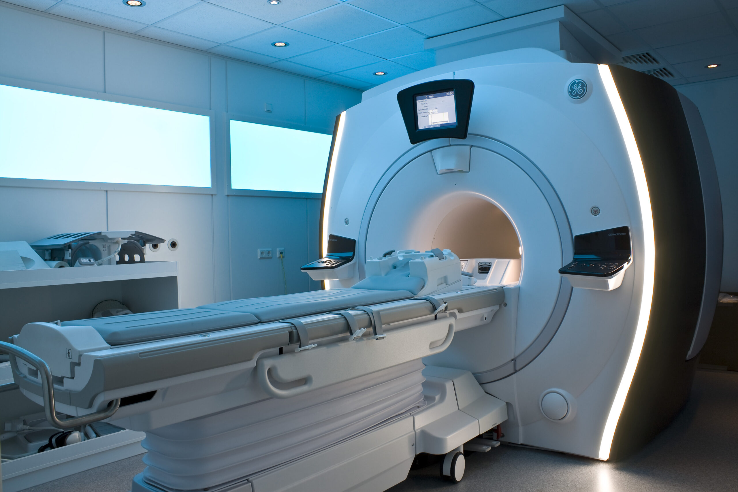
To appreciate the functionality of an MRI machine, it is essential to understand its operational principles. The apparatus consists of a large magnet, typically a superconducting magnet, which produces a strong and uniform magnetic field. When a patient is positioned within this magnetic field, the protons in the body—predominantly in water and fat—align with the direction of the magnetic field. Subsequently, radiofrequency pulses are administered, which temporarily disturb this alignment. As the protons return to their equilibrium state, they emit radiofrequency signals that are captured and processed to create detailed cross-sectional images of the anatomy.
This interaction between magnetic fields and radio waves can be likened to a symphony, where the magnetic field serves as the conductor, coordinating the notes emitted by the protons. The resulting images vary in contrast based on the tissue composition, yielding distinctions that enable clinicians to discern between healthy and diseased states. T1 and T2 relaxation times, intrinsic properties of the tissues, facilitate various imaging sequences, further enhancing the diagnostic capabilities of MRI.
The clinical applications of MRI are extensive and multifaceted. One of the most prolific uses is in neuroimaging, where MRI excels in delineating the structure and function of the brain and spinal cord. Conditions such as tumors, multiple sclerosis, and cerebrovascular accidents are meticulously evaluated using MRI. In addition, musculoskeletal MRI has revolutionized the assessment of joint and soft tissue pathology, enabling the identification of injuries that may be elusive on other imaging modalities.
Moreover, MRI plays a critical role in oncological imaging, where it assists in tumor characterization and staging. The ability to visualize the extent of cancerous growth and its proximity to surrounding structures enhances surgical planning and treatment protocols. Furthermore, functional MRI (fMRI) offers a remarkable dimension, allowing researchers and clinicians to observe brain activity by measuring changes in blood flow, fundamentally altering our understanding of neurological processes.
As MRI technology continues to evolve, innovative approaches such as diffusion tensor imaging (DTI) and spectroscopy are augmenting standard imaging techniques. DTI, for instance, provides insights into neural pathways and connectivity within the brain, thereby heralding a new era in the study of neurodegenerative diseases and psychiatric disorders. Spectroscopy allows for the assessment of biochemical changes within tissues, granting valuable information regarding metabolic processes, which could be paramount in cancer diagnosis and treatment monitoring.
The resolution and speed of MRI imaging are also poised to benefit from advancements in hardware technology, including the advent of high-field MRI systems that operate at magnetic strengths greater than 3 Tesla. These systems promise unparalleled image clarity and detail, significantly enhancing diagnostic accuracy. Moreover, the development of robust algorithms for image reconstruction is facilitating faster scan times, minimizing patient discomfort and enhancing throughput in clinical settings.
Furthermore, artificial intelligence is permeating the realm of MRI, providing unprecedented opportunities for improving diagnostic workflows. Machine learning algorithms are increasingly being trained to analyze imaging data, detect anomalies, and predict outcomes with remarkable accuracy. The integration of AI into MRI not only streamlines the diagnostic process but also augments the decision-making capabilities of radiologists and clinicians.
Despite these advancements, the implementation of MRI technology is not without its challenges. The high cost of MRI machines and the necessity for specialized training for operators can serve as barriers to access, particularly in resource-limited environments. Furthermore, patients with certain implants, such as pacemakers, may be ineligible for MRI scans due to potential interactions with the magnetic field. Addressing these challenges is essential to ensure that the benefits of MRI technology can be realized universally across different populations.
In summary, MRI represents a remarkable confluence of physics and medicine, epitomizing the possibilities that arise from meticulous scientific inquiry. Its capacity to non-invasively visualize internal structures and processes has irrevocably altered the landscape of diagnostic imaging. With ongoing technological advancements, the promise of MRI is set to expand dramatically, opening new frontiers in medical science and patient care. As we delve deeper into the complexities of the human body through MRI, we move closer to uncovering the mysteries that lie within, propelling us toward a more nuanced understanding of health and disease.
