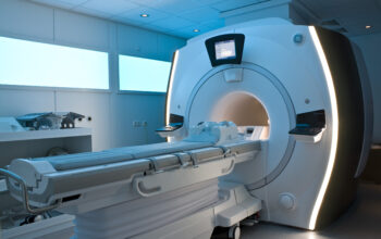
Diagnostic medical sonography, often referred to simply as ultrasound, represents a vital intersection of imaging technology and clinical practice. It serves critical roles in various medical contexts, providing both diagnostic and therapeutic capabilities. However, despite its prevalence, many may wonder: can the complexities of human anatomy truly be visualized in real-time, using nothing more than sound waves? This inquiry leads to an exploration of the multifaceted discipline that challenges our understanding of both technology and medicine.
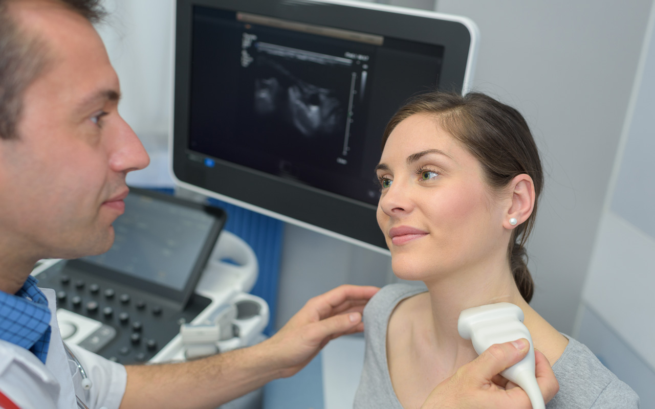
At its core, diagnostic medical sonography employs high-frequency sound waves to produce images of the internal structures of the body. These sound waves, emitted by a transducer, penetrate tissue and reflect back, forming a sonographic image. This non-invasive technique is safe, devoid of the ionizing radiation risks associated with X-rays or CT scans, thus making it a preferred choice in various clinical settings.
One of the most captivating attributes of sonography is its versatility. It finds applications across a spectrum of medical fields, including obstetrics, cardiology, and abdominal imaging. In obstetrics, sonography allows for the visualization of fetuses in utero, aiding in the assessment of fetal health and development. As a playful question: could the intricate details of early life development truly be captured in such a straightforward, safe manner? The answer reveals the remarkable capability of ultrasound technology.
In cardiovascular diagnostics, sonography can evaluate heart structure and function through echocardiograms. This modality enables clinicians to visualize the heart’s chambers, valves, and blood flow dynamics, providing insights into various cardiac conditions. Moreover, Doppler ultrasound techniques can measure the speed of blood flow, further enhancing diagnostic accuracy.
Beyond obstetrics and cardiology, diagnostic medical sonography is invaluable in assessing abdominal organs, such as the liver, gallbladder, kidneys, and pancreas. Understanding the anatomy and pathology of these organs through sonographic evaluation can significantly impact patient management. The question thus arises: how can such detailed insights be gleaned without invasive procedures?
In addressing the array of conditions that sonography can detect, several noteworthy applications are often highlighted. Conditions such as gallstones, liver cirrhosis, kidney stones, and even pancreatic tumors can be diagnosed using ultrasound. Ultrasound may also assist in guiding biopsies, aiding interventions, and monitoring disease progression through serial imaging. This non-invasive nature poses both a profound benefit for patient comfort and a challenge for diagnostic accuracy.
Understanding the technical aspects of sonography is equally compelling and essential. The process begins with the transducer, which functions as both a transmitter and a receiver of ultrasound waves. The interaction between sound waves and tissue varies according to the density and composition of the material. This interaction forms the basis of echogenicity, which refers to the ability of tissues to reflect ultrasound waves. Structures such as bone and air produce high echogenicity, appearing bright on ultrasound images, while fluid-filled structures, like cysts, appear dark.
Nevertheless, the interpretation of sonographic images is where the true challenge lies. The operator’s skill and experience play a critical role in accurately diagnosing conditions. Artifacts, which are unintended echoes or shadows on the ultrasound images, can obscure true anatomical structures, thus complicating interpretation. Moreover, various factors such as patient anatomy, body habitus, and the skill of the sonographer can influence image quality. Thus, a question of expertise arises: is artistic perception on par with scientific knowledge when deciphering the nuances of ultrasound images?
The realm of diagnostic medical sonography is not without its limitations and challenges. One of the more pronounced drawbacks is the inability to penetrate through bones or gas-filled structures, thereby limiting the visualization of certain anatomical areas. Additionally, the quality of the images can be compromised in obese patients due to increased tissue thickness, necessitating a combination of imaging modalities for comprehensive evaluation in such cases.
As the field of diagnostic medical sonography matures, advancements in technology continue to amend these limitations. Innovations such as three-dimensional (3D) and four-dimensional (4D) ultrasound have emerged, enhancing the diagnostic capabilities of traditional techniques. Such advancements allow for volumetric analysis of structures, facilitating more detailed assessments of fetal development in obstetrics and refining the understanding of complex anatomical relationships in other areas of the body.
Continuing education and training for sonographers also play a pivotal role in advancing the field. As technology evolves, the need for skilled professionals who can adeptly navigate these innovations becomes increasingly paramount. Moreover, fostering interdisciplinary collaboration among radiologists, obstetricians, and other healthcare professionals ensures that patients benefit from a comprehensive perspective on their diagnosis and treatment.
In conclusion, diagnostic medical sonography embodies a crucial aspect of modern medicine, offering a non-invasive, safe, and flexible imaging modality that enriches our understanding of the human body. As sophisticated technologies evolve, one might ponder: how far can our exploration of the physiological human landscape extend through the serene echoes of sound? The interplay between technological advancement, clinical acumen, and educational initiatives will undoubtedly shape the future trajectory of this indispensable field, cementing its role in diagnostic medicine. This is a dynamic space, ripe for exploration, innovation, and the continual unraveling of medical mysteries through the power of sound.