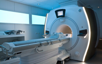
Magnetic Resonance Imaging (MRI) has revolutionized the field of diagnostic imaging, enabling clinicians to visualize intricate anatomical structures and pathological conditions with unprecedented detail. Within an MRI scan, various parameters are adjusted to optimize the imaging of specific tissues or conditions. Among these parameters, three terms are frequently encountered: ST (Short Tau), ET (Echo Time), and RT (Repetition Time). In this article, we will delve into these terms, elucidating their definitions, clinical significance, and implications for MRI interpretation.
Understanding MRI Fundamentals
Before examining ST, ET, and RT, it is imperative to grasp the fundamental principles governing MRI technology. MRI exploits the magnetic properties of hydrogen nuclei, predominantly found in water and fat within the human body. When subjected to a strong magnetic field, these nuclei align with the magnetic field lines. A pulse of radiofrequency energy is then applied, which excites the protons, causing them to resonate and subsequently re-emit energy as they return to their equilibrium state. This emitted signal is captured and transformed into images. The timing of these pulses is crucial in determining the appearance of the resulting images.
1. Repetition Time (RT)
Repetition Time (RT) refers to the time interval between successive radiofrequency pulses applied to the same slice of tissue. It is a fundamental parameter that influences image contrast and tissue differentiation in MRI scans. RT is measured in milliseconds (ms) and directly impacts the relaxation processes of protons. Specifically, it affects T1 relaxation, which relates to the time it takes for protons to return to their longitudinal alignment after excitation.
Short RT values tend to emphasize T1-weighted images, making fat-containing tissues appear bright, whereas fluids such as cerebrospinal fluid may appear darker. Conversely, long RT values enhance T2-weighted images, wherein fluid-filled structures are vividly displayed. The choice of RT is therefore pivotal, depending on the clinical question at hand. For example, evaluating brain edema may require a longer RT for optimal visualization of fluid accumulation, while assessing liver lesions may necessitate shorter RT values to distinguish between different tissue types.
2. Echo Time (ET)
Echo Time (ET) denotes the time elapsed between the application of the radiofrequency pulse and the receipt of the emitted signal. This interval is crucial in defining the degree of T2 contrast in the resultant images. Similar to RT, ET is also expressed in milliseconds and serves as a key determinant of image quality and diagnostic efficacy.
Short ETs are suitable for generating T1-weighted images, which emphasize fat-rich tissues by optimizing the visualization of T1 recovery processes. In contrast, longer ET values enhance T2-weighted images, facilitating the imaging of liquids. For instance, in cases of tumors or lesions containing significant fluid, a longer ET can accentuate the pathological features that would otherwise be missed with shorter ETs.
Additionally, ET can be adjusted in various imaging sequences to optimize the visualization of different tissues. For example, in a proton density-weighted sequence, a balanced ET is employed to capture the density of protons across different tissues, allowing for a distinct evaluation of fibrous tissues or cartilage.
3. Short Tau (ST)
Short Tau (ST), albeit less commonly referenced than RT and ET, plays a crucial role in advanced imaging techniques such as diffusion-weighted imaging (DWI) or contrast-enhanced MR imaging. The term “tau” refers to the timing of the inversion pulse in these sequences. In the context of ST, it relates to the timing of a specific inversion pulse applied to create a specific tissue contrast that is sensitive to specific pathological processes.
In certain sequences, such as in MR angiography, ST can delineate vascular structures with high precision by exploiting the timing of echo acquisition relative to blood flow. It is particularly useful in visualizing perfusion abnormalities or assessing vascular malformations. Consequently, optimizing ST is critical for detecting conditions that may otherwise evade simple structural assessment.
Clinical Implications of RT, ET, and ST
The interplay of Repetition Time, Echo Time, and Short Tau significantly impacts the diagnostic capacity of MRI. The selection of appropriate parameters can refine image quality, enhance tissue contrast, and improve the delineation of pathologies. Understanding these parameters is essential for radiologists and medical professionals for accurate image interpretation and effective clinical decision-making.
Moreover, these timing sequences must be tailored for specific indications. For instance, in neuroimaging, RT and ET can be adjusted to optimize the assessment of brain lesions, while in musculoskeletal imaging, precise manipulation of these variables can aid in revealing subtle cartilage injuries or muscle tears. Ultimately, an appreciation of RT, ET, and ST enhances diagnostic accuracy and fosters effective patient care through tailored imaging techniques.
Conclusion
In summary, RT, ET, and ST are critical parameters that govern the quality and diagnostic utility of MRI scans. Their meticulous adjustment allows for the optimization of image contrast, enabling clinicians to differentiate between various tissue types and identify pathological conditions with heightened clarity. As MRI technology continues to evolve, a robust understanding of these principles will remain indispensable in guiding clinical practices and advancing patient outcomes.

