The evolution of imaging technologies, particularly in the realm of radiography, has profoundly transformed diagnostic practices. Among these innovations, the advent of dark field imaging represents a significant paradigm shift in our understanding and application of X-ray technology. But what happens when we illuminate the shadowy realms of conventional imaging techniques? Is it possible that by shifting our perspective and utilizing advanced methodologies, we can unearth a wealth of previously obscured diagnostic information?
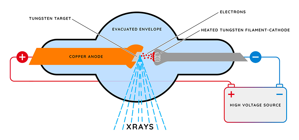
To appreciate the implications of dark field imaging, we must first comprehend the principles governing traditional X-ray imaging. In standard applications, X-rays penetrate tissues of varying densities and, depending on their atomic composition, create a radiograph that reveals contrasts in density as varying shades of black and white. However, this conventional method is frequently limited by its diagnostic capacity, particularly when examining soft tissues or detecting subtle pathologies.
Dark field imaging diverges from the traditional paradigm by taking advantage of the principles of scatter and reflection. Rather than merely capturing the direct transmission of X-rays through a specimen, dark field techniques assess the weakly scattered X-rays—a phenomenon that yields valuable information about the microstructural characteristics of the imaged object. By utilizing a specialized detector configuration, dark field imaging can highlight contrasts that would otherwise remain imperceptible, thus enhancing the diagnostic yield in countless clinical scenarios.
The core advantage of dark field imaging lies in its ability to visualize low-contrast features, particularly within soft tissues where conventional X-ray modalities tend to falter. For instance, entities such as tumors or micro-calcifications may elude identification under standard conditions, yet they often exhibit specific scattering patterns that dark field techniques can illuminate. By focusing on light that is scattered at angles greater than a predetermined threshold, the images produced not only enhance visibility of the target structures but also encapsulate essential information concerning their morphology and spatial distribution.
Despite the advantages celebrated by the proponents of dark field imaging, these novel methods are not without their challenges. The transition from a well-established imaging domain to a more sophisticated and somewhat esoteric technique can provoke skepticism among practitioners. This leads to an intriguing question: how do we reconcile the innovative potential of dark field imaging with the entrenched practices that dominate medical imaging today? The potential challenge resides in the necessity to educate and inform radiologists about not just the capabilities but also the integration of these advanced techniques into routine clinical workflows.
Indeed, empirical studies have begun to substantiate the efficacy of dark field imaging across varied applications. Research endeavors have demonstrated the method’s efficacy in musculoskeletal imaging, where subtle fractures or anomalies obscured by overlying structures can be made apparent. In pulmonary imaging, dark field techniques have opened avenues for the detection of early-stage lung disease, thereby averting the progression to more advanced, often less treatable conditions.
The integration of dark field imaging into clinical practice also raises pivotal questions about cost-effectiveness and accessibility. As health systems strive to deliver value-based care, the question of whether the potential gains in diagnostic accuracy can render these advanced modalities economically feasible remains salient. Moreover, should these techniques necessitate substantial investment in new equipment or extensive training, one must ponder whether the healthcare landscape is prepared for such a transformative shift.
Furthermore, the advent of artificial intelligence (AI) and machine learning holds incredible promise for advancing the capabilities of dark field imaging. As algorithms become increasingly adept at interpreting intricate and ambiguous datasets, they can enhance the accuracy and reliability of interpretations. This begs the question of a new frontier: will the fusion of dark field imaging and AI lead to an unprecedented dimension of diagnostic precision, or will it provoke ethical dilemmas related to the dependency on technology over human expertise?
As the exploration of dark field imaging progresses, ongoing interdisciplinary collaboration will be paramount. Radiologists, physicists, and technologists must unite to share insights, troubleshoot challenges, and glean new perspectives. This collaborative approach will not only elevate the understanding of dark field modalities but also foster the refinement and application of these methods in diverse diagnostic contexts.
Dark field imaging emerges as a powerful tool that sheds light on the hidden intricacies of our biological structures. The capacity to elucidate otherwise imperceptible details creates opportunities for more precise diagnostics and enhanced patient outcomes. Yet, the interplay between innovation and established practice poses as both a catalyst for advancement and a formidable challenge to navigation.
In summation, the evolution of dark field imaging amongst the various modalities of X-ray techniques encapsulates a forward-thinking trajectory in diagnostic radiography. It challenges us to consider two cardinal principles: the potential lying within unexplored frameworks and the necessity of meticulous integration into practice. Thus, addressing the interplay of skepticism, cost, and technological advancement is imperative as we forge ahead, illuminating the darker corners of diagnostic imaging from which valuable information can ultimately emerge.