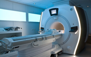
Magnetic Resonance Imaging (MRI) is a powerful diagnostic tool that has revolutionized our understanding of human anatomy and physiology. However, while MRI is widely recognized for its ability to generate detailed images of soft tissues within the body, the question of what kind of light an MRI emits might seem perplexing at first glance. To comprehend the interaction between MRI technology and light emission, one must delve into the principles underlying both magnetic resonance and the electromagnetic spectrum.
At its core, MRI capitalizes on the principles of nuclear magnetic resonance (NMR). This technique exploits the magnetic properties of atomic nuclei, primarily hydrogen, to derive high-resolution images of internal body structures. The emitted signals from the hydrogen nuclei, excited by a strong magnetic field, do not include light in the visible spectrum but are instead associated with specific radiofrequency (RF) emissions. Thus, when contemplating the “light” of an MRI, it is crucial to clarify that we are referring to RF waves, which lie at the lower frequency end of the electromagnetic spectrum.
Understanding the nature of these radio waves necessitates an exploration of the electromagnetic spectrum itself. The spectrum is typically categorized into a range of wavelengths and frequencies, from gamma rays at one end through to radio waves at the other. The radiofrequencies employed in MRI are typically in the range of 10 to 100 megahertz (MHz), which is substantially lower than visible light, infrared radiation, or ultraviolet light. The differential characteristics of these electromagnetic waves determine their utility in medical imaging. While gamma rays can penetrate through tissue, and x-rays are adept at delineating structured density, MRI utilizes RF emissions due to their non-ionizing nature, rendering them safer for patient use.
Within the confines of MRI technology, the role of RF pulses is paramount. These pulses serve as exogenous perturbations that disrupt the alignment of hydrogen nuclei within a strong magnetic field. When these spins relax back to equilibrium, they emit RF signals that can be detected and translated into high-fidelity images. The characteristics of these emitted signals include various parameters such as T1 and T2 relaxation times, which reflect different tissue properties. Herein lies the intersection of physics and medical diagnostics; the manipulation and analysis of electromagnetic waves yield insights that are pivotal for clinical evaluation.
Different MRI sequences can yield distinct types of images, including T1-weighted, T2-weighted, and proton density-weighted images, each highlighting varying aspects of tissue characteristics. The modulation of RF pulses, along with gradient fields, enables the resolution of minute anatomical structures and pathologies. Each sequence can arguably reveal a different ‘face’ of the same underlying physiology, being contingent on the timing of pulse sequences and slicing techniques utilized.
One must also consider the phenomenon of induced magnetism, which interplays with the RF emissions in MRI technology. During magnetic resonance, the hydrogen nuclei become polarized in the magnetic field. These nucleuses exhibit a net magnetic moment, and during the application of RF pulses, they resonate. This resonance results in the generation of secondary electromagnetic waves, which are in essence the ‘light’ emitted during an MRI scan. However, it is a misnomer to equate these emissions with visible light; rather, they contribute to a complex diagnostic imaging process devoid of photonic emission in the visible spectrum.
Furthermore, the field of MRI is constantly evolving, integrating advanced techniques such as functional MRI (fMRI) and diffusion tensor imaging (DTI). Functional MRI employs RF signals to measure brain activity by detecting changes in blood flow, correlating with neural activity. The integration of optical techniques with MRI, such as the aforementioned novel sensor technology that utilizes MRI to detect light, represents a burgeoning area of research. This interdisciplinary innovation underscores the potential emergence of optical imaging modalities that could complement traditional MRI functionality.
Importantly, while RF emissions are the primary exponents of the diagnostic capability of an MRI machine, various safety concerns arise regarding electromagnetic exposure. Although RF waves are non-ionizing, it remains essential to monitor exposure levels to mitigate potential thermal effects that can arise from prolonged imaging sessions. As research continues into the long-term implications of exposure, regulatory guidelines are adapted to enhance patient safety and operational efficacy.
In conclusion, while an MRI machine may not emit light in the conventional sense, understanding its output encompasses a grasp of radiofrequency emissions and their extraordinary capabilities. The intricacies of the electromagnetic spectrum and the fundamental principles of physics underpinning MRI technology are indispensable for appreciating its impact on modern medicine. Through continuous advancements and interdisciplinary approaches, the capability of MRI to elucidate the complexities of human biology will undoubtedly expand, fostering enhanced diagnostic prowess and therapeutic interventions.


