Nuclear Magnetic Resonance (NMR) imaging, commonly known as Magnetic Resonance Imaging (MRI), stands as a revolutionary non-invasive diagnostic tool predominantly utilized in medical imaging to visualize the internal structures of the human body. This sophisticated imaging technique harnesses the principles of nuclear magnetic resonance to produce detailed anatomical maps of soft tissues. Understanding the theoretical underpinnings, technological advancements, and clinical applications of NMR imaging can illuminate its critical role in contemporary medicine.
At the core of NMR imaging lies the concept of nuclear magnetic resonance, wherein certain atomic nuclei are subjected to an external magnetic field, leading to the absorption and emission of electromagnetic radiation. Hydrogen nuclei, abundantly present in biological tissues due to high water content, are primarily utilized in NMR. When exposed to a magnetic field, these nuclei align with the field and subsequently undergo precession. When subjected to radiofrequency pulses, they absorb energy, transitioning to higher energy states. This energy is released upon relaxation, during which the nuclei return to equilibrium, producing signals that can be detected and translated into images by sophisticated software.
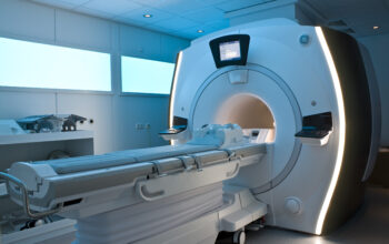
The operational mechanism of NMR imaging involves several critical components: the magnet, radiofrequency coil, and gradient coils. The magnet generates a sustenance magnetic field, typically measured in teslas (T). Clinical MRI systems commonly operate at 1.5T or 3T. High-field magnets yield higher signal-to-noise ratios, fostering improved image clarity at the cost of accessibility and affordability. The radiofrequency coil excites the hydrogen nuclei, while the gradient coils manipulate the magnetic field strength, enabling spatial localization of the emitted signals. These components collectively facilitate the generation of high-resolution images and the differentiation of various tissue types based on their specific magnetic properties.
Advanced techniques have emerged within the realm of NMR imaging, enhancing the versatility of this modality. Functional MRI (fMRI), for instance, leverages the hemodynamic response, where neuronal activity correlates with blood flow changes. This mechanism enables the visualization of brain activity during specific cognitive tasks, significantly contributing to neuroscience and psychology. Diffusion-weighted imaging (DWI) is another salient development that evaluates the diffusion of water molecules within tissues. This technique is particularly valuable in the assessment of stroke, as it can highlight areas of restricted diffusion indicative of ischemic conditions.
The resolution of anatomical structures achieved through NMR imaging is profound. Viewer engagement with such high-fidelity images reveals the intricate details of organs, tissues, and even pathological changes. For example, the use of contrast agents—substances introduced into the body to enhance the visibility of specific tissues—further amplifies diagnostic capability. Gadolinium-based contrast agents are widely administered, allowing for enhanced observation of vascular structures, tumors, and inflammatory processes. Yet, it is imperative to acknowledge the consideration of potential nephrotoxicity and contraindications in patients with renal insufficiency.
The clinical applications of NMR imaging are vast, spanning various medical disciplines. Neurology extensively utilizes MRI for the diagnosis and management of conditions such as multiple sclerosis, brain tumors, and traumatic brain injuries. Oncologists rely on MRI for tumor characterization, staging, and treatment monitoring, enabling tailored therapeutic strategies based on precise tumor localization and morphology. In orthopedics, MRI excels at visualizing soft tissue injuries, ligaments, and cartilage, providing invaluable insights into musculoskeletal disorders.
Despite its immense capabilities, certain limitations and challenges accompany NMR imaging. For one, the technique is inherently sensitive to motion artifacts, which can obscure diagnostic accuracy. Patients who are unable to remain still, such as young children or individuals suffering from anxiety, may present challenges during scanning procedures. Additionally, the relatively extended duration of MRI scans compared to other imaging modalities, such as computed tomography (CT), can lead to discomfort and claustrophobia among patients. Furthermore, accessibility remains a concern, as MRI machines are costly and often limited in availability in certain healthcare settings.
Ongoing research is paramount in addressing these challenges and advancing NMR imaging technology. The development of higher field strength magnets, faster imaging sequences, and novel contrast agents is at the forefront of innovation. Moreover, efforts to integrate artificial intelligence (AI) into the image acquisition and interpretation processes promise to enhance diagnostic accuracy and streamline workflows, potentially reducing the burden on radiologists.
In summation, NMR imaging, grounded in the principles of nuclear magnetic resonance, has emerged as a cornerstone of modern diagnostic radiology. Its capacity to produce high-resolution images non-invasively makes it an invaluable asset in numerous clinical settings. With continued advancements in the technology, there lies a tantalizing prospect for enhancing both diagnostic precision and patient care. As the field evolves, embracing the intricate interplay of physics, technology, and biology will serve to unlock the full potential of this remarkable imaging modality.


