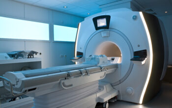
Magnetic Resonance Imaging (MRI) has become an indispensable tool in contemporary medical diagnostics, offering an advanced imaging modality that provides detailed anatomical and physiological information without the use of ionizing radiation. The primary question of interest is encapsulated in the term itself: what does the ‘M’ in MRI signify? This inquiry delves into the fundamental principles and innovative technology underpinning MRI, as well as the diverse applications and implications of this radiological technique.
The ‘M’ in MRI stands for “Magnetic,” which is a reference to the technology’s reliance on powerful magnets as the core component of its imaging methodology. To understand the significance of magnetism in this context, it is essential to elucidate the principles of nuclear magnetic resonance (NMR). The phenomenon is predicated on the magnetic properties of atomic nuclei, specifically the hydrogen nuclei (protons) that are abundant in biological tissues due to the high-water content of the human body. When individuals are placed within a strong magnetic field, the protons in their bodies align with this field, creating a state of magnetization.
The operational principles of MRI involve not merely the application of magnetism; rather, they necessitate the precise manipulation of radiofrequency (RF) pulses. These RF pulses are employed to perturb the alignment of the protons, prompting them to resonate. Once the RF pulse is turned off, the protons gradually return to their original alignment within the magnetic field. This reassignment process emits signals, which are subsequently detected by the MRI machine and transformed into images through complex algorithms. As such, the intricate interplay between magnetism, radiofrequency energy, and signal processing lies at the heart of MRI technology.
There are various types of MRI scans that cater to different diagnostic needs, emphasizing the versatility of this imaging technique.
- Functional MRI (fMRI): This variant focuses on detecting brain activity by measuring changes in blood flow. fMRI exploits the principle of cerebral oxygenation, providing insights into metabolic activity, cognitive functions, and potential abnormalities in brain structure and function.
- Cardiac MRI: Specialized for heart imaging, cardiac MRI offers a detailed view of cardiac anatomy and function, allowing for the assessment of myocardial perfusion, structural anomalies, and the evaluation of cardiomyopathies. This type of MRI employs tailored imaging sequences to capture real-time cardiac motion.
- Magnetic Resonance Angiography (MRA): MRA is specifically designed to visualize blood vessels. By utilizing contrast agents or advanced imaging techniques, MRA facilitates the examination of vascular diseases, aneurysms, and stenosis without the need for invasive procedures.
- Diffusion Tensor Imaging (DTI): An advanced form of MRI, DTI assesses the movement of water molecules in biological tissues, particularly within the brain’s white matter. This modality is crucial for studying neural pathways and is employed in understanding traumatic brain injuries and neurodegenerative diseases.
The implications of MRI extend beyond mere imaging capabilities; it revolutionizes diagnostic protocols and therapeutic planning across various specialties. For instance, in oncology, MRI elucidates tumor characterization, staging, and response to therapy, thereby guiding treatment regimens. Similarly, in the realm of neurology, MRI is instrumental in diagnosing conditions such as multiple sclerosis, stroke, and brain tumors, providing invaluable information that influences management strategies.
However, the application of MRI is not devoid of considerations and challenges. The magnetic fields employed in MRI introduce particular safety concerns. Patients with ferromagnetic implants or devices, such as pacemakers, face contraindications due to the potential for device malfunction or displacement. Furthermore, the claustrophobic nature of traditional MRI scanners often poses challenges for patients. Newer technologies, such as open MRI systems, have emerged to mitigate these discomforts, although they may compromise image resolution.
In addition to the logistical challenges, the interpretation of MRI scans necessitates significant expertise. Radiologists must possess a comprehensive understanding of normal and pathological anatomy, alongside familiarity with various imaging techniques and artifact recognition. The assimilation of advanced machine learning and artificial intelligence into MRI interpretation is proving advantageous, enhancing diagnostic accuracy and potentially expediting clinical workflows.
In a broader context, the future of MRI technology appears promising. Research continues to advance the boundaries of MRI applications, including the integration of molecular imaging, enhanced resolution techniques such as ultra-high-field MRI, and the potential for real-time imaging capabilities. These innovations may further revolutionize the capabilities of MRI, culminating in improved patient outcomes and expanded diagnostic horizons.
In summary, the ‘M’ in MRI encapsulates the fundamental principle of magnetism central to this sophisticated imaging technique. From its foundational physical principles to its diverse applications across numerous medical fields, MRI serves as a testament to the synergy between technology and healthcare. As innovations in both hardware and software continue to evolve, the realm of MRI is poised to enhance its status as a cornerstone of modern diagnostics, ultimately enriching patient care and medical knowledge.


