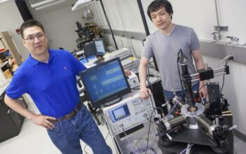
As the frontiers of fundamental science push ever deeper into the nanoworld, electron tomography emerges as an invaluable tool, unlocking the intricate architecture of materials and biological specimens at resolutions previously deemed unattainable. This multidimensional analytical technique combines the principles of electron microscopy with tomographic reconstruction, enabling researchers to visualize structures that reside within the sub-100 nanometer regime. This article delves into the fundamentals of electron tomography, its myriad applications, and the transformative insights it affords to the scientific community.
Electron tomography is fundamentally the application of electron microscopy to acquire a series of two-dimensional projection images from multiple angles around a specimen. These images are subsequently reconstructed into a three-dimensional representation, offering an unparalleled glimpse into the internal configurations of specimens. The advent of this technique marked a significant milestone in materials science, biology, and nanotechnology, where understanding spatial arrangements and architecture is crucial for deciphering functional properties.
The precision of electron tomography hinges predominantly on two methodologies: single-particle analysis and cryo-electron tomography (cryo-ET). Each of these methodologies serves distinct yet complementary purposes in the exploration of materials and biological entities at the nanoscale. Single-particle analysis, for instance, predominantly focuses on isolated macromolecules or complexes, elucidating structural characteristics with phenomenal clarity. This method is particularly advantageous in structural biology, where it elucidates the architecture of proteins, viruses, and larger biomolecular assemblies.
In contrast, cryo-electron tomography offers the ability to visualize frozen-hydrated specimens in their native environments. This technique eliminates the artifacts associated with sample preparation, allowing researchers to observe cellular structures and organelles as they exist in vivo. By subjecting samples to rapid cryogenic freezing, cellular dynamics can be retained and studied, providing crucial insights into the interaction of biomolecules within cellular compartments. This methodology has profound implications for the study of cellular processes, including endocytosis and mitochondrial dynamics, thereby enhancing our understanding of life at the molecular level.
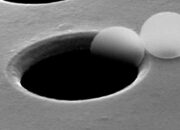
The process of acquiring tomographic data involves meticulous attention to detail and precision. Typically, this begins with the selection and preparation of the specimen, which must be thin enough to allow electrons to penetrate. Sample thinning can be achieved through various methods, including focused ion beam milling and ultramicrotomy. Once prepared, the specimen is placed in an electron microscope capable of tilt-series imaging, where it is incrementally rotated, capturing numerous projection images from distinct angles.
The subsequent reconstruction of these images requires advanced computational algorithms tailored for tomographic reconstruction. The most widely employed method is the weighted back-projection technique, which reconstructs the three-dimensional structure by mathematically combining the projection images. As computation technology has advanced, so too has the efficacy of these algorithms, allowing for improved resolution and reduced noise in the final tomographic volumes.
Electron tomography is not confined to purely academic pursuits. The implications of this technology extend to a myriad of industries, including semiconductor manufacturing, nanotechnology, and pharmaceuticals. In semiconductor manufacturing, for instance, electron tomography facilitates the inspection of microelectronic components at the nanoscale, allowing engineers to identify defects and optimize designs. Such insights are critical, as the drive towards miniaturization necessitates impeccable precision at the atomic level.
The pharmaceutical industry also benefits profoundly from the ability to visualize drug-target interactions at the molecular level. By elucidating the conformational states of proteins in the presence of ligands, researchers can gain insights into mechanism-of-action pathways, enabling the design of more effective therapeutics. Moreover, as personalized medicine becomes increasingly prevalent, understanding the structural nuances of biomolecules in specific patient profiles becomes paramount, a task electron tomography is well-positioned to address.
Despite its myriad advantages, challenges remain in the application of electron tomography. One notable limitation is related to the inherent noise and artifacts that can obscure structural details, particularly when dealing with heterogeneous samples. Ongoing research is focused on ameliorating these issues through advancements in imaging techniques and computational methods. Additionally, the high demands placed on sample preparation and the sophisticated technology required for data acquisition and processing mean that access to electron tomography resources is often confined to specialized research centers and institutions.
Looking forward, the future of electron tomography appears promising, enabled by emerging technologies such as machine learning and artificial intelligence. These advancements hold the potential to streamline data processing, enhance image reconstruction, and automate aspects of data acquisition. Furthermore, as techniques like cryo-electron tomography continue to mature, the scope of applicability will undoubtedly expand, fostering interdisciplinary collaborations that span the realms of physics, biology, and materials science.
In conclusion, electron tomography stands at the vanguard of nanoscale research, offering an intricate view into the structural tapestry of materials and biological specimens. With its ability to transcend the boundaries of traditional microscopy, it fosters a deeper understanding of the fundamental processes governing the natural world. The ongoing evolution of this technique promises to unveil further mysteries of the nanoworld, propelling scientific inquiry into new and uncharted territories.