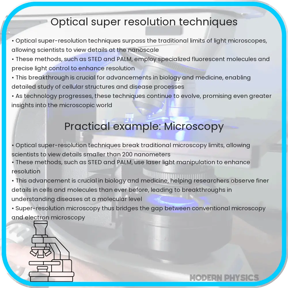
In the realm of modern physics, the pursuit of optical super-resolution stands as a resounding testament to human ingenuity—a symphony where light and matter dance in intricate harmony. The quest for surpassing the diffraction limit—an omnipresent boundary that confines the resolution of optical microscopy—has galvanized researchers and innovators alike. Innovative techniques have emerged that defy conventional knowledge, illuminating the vast potential of the microscopic world.
The fundamental principle of optical imaging rests upon the behavior of light as it interacts with matter, a relationship marred by the constraints imposed by diffraction. At its core, diffraction acts as an insurmountable barrier, akin to a foggy veil that obscures the true essence of minuscule entities. The resolution limit, traditionally pegged at approximately half the wavelength of light, confines the observer to a world seen through murky glass, preventing the exquisite details of cellular structures and molecular interactions from being fully appreciated.
To breach this veil, researchers have embraced a plethora of innovative methodologies, varying in elegance and complexity, yet unified by a common goal: to unveil the microcosm with unparalleled precision. Techniques such as Stimulated Emission Depletion (STED) microscopy, Single-Molecule Localization Microscopy (SMLM), and the ingenious applications of Structured Illumination Microscopy (SIM) have emerged as pioneering nodes in this extraordinary journey.
STED microscopy epitomizes the ingenuity of optical engineering. In this technique, an additional laser beam is employed to selectively deplete fluorescence from all but a tantalizingly small area, thus sharpening the focus of the imaging process. This is reminiscent of a sculptor chiseling away extraneous material to reveal a portrait hidden within marble. The ability to manipulate light in such a manner not only enhances resolution but elucidates dynamic biological processes with unprecedented clarity.
Equally revolutionary is the approach embodied in SMLM, which capitalizes on the stochastic activation of fluorescent molecules. This method hinges on the principle that by observing individual fluorescent molecules over time, one can reconstruct a high-resolution image through computational techniques. Each molecule acts as a point source of illumination, akin to the scattered stars in a midnight sky, collectively forming constellations of molecular interactions that have been previously obscured. The application of this method grants researchers the capability to explore cellular structures at the nanometer scale, illuminating the previously hidden architecture of biological systems.
Another noteworthy innovation, Structured Illumination Microscopy (SIM), employs patterned illumination to comprehend otherwise complicated three-dimensional biological samples. By projecting intricate light patterns onto specimens, SIM compels the underlying structures to reveal themselves in sharp relief. This innovative interplay between light and structure cultivates an understanding of intricate biological relationships. The technique propels researchers into a realm where they can visualize the choreography of molecular interactions with astounding clarity.
Each of these methodologies not only enhances resolution but dramatically transforms the landscape of biological research, where the study of live cells is no longer relegated to a mere representation of life but is instead an engaging exploration of the living world in real-time. The challenge of managing the overwhelming data generated by these techniques presents a daunting yet exhilarating frontier. The sheer volume of information, the sparkle of countless molecular symphonies happening simultaneously, requires robust computational tools to distill meaning from the noise.
Machine learning has emerged as a bastion of sophistication in this context. Integrating multiscale data analysis with image reconstruction enables the refinement of captured data, ultimately achieving a new zenith in imaging resolution and interpretative power. As algorithms learn to recognize patterns and extract information seamlessly, they become invaluable accomplices in the effort to illuminate the intricate behaviors of cells, proteins, and other biological constituents. The future appears fertile with potential as the intersection of optical super-resolution and machine learning continues to burgeon.
Furthermore, the implications of optical super-resolution extend far beyond the confines of biology and microscopy. In fields ranging from materials science to nanotechnology, the ability to resolve structures and phenomena at the nanoscale catalyzes profound advancements. Innovations in nanomaterials, drug delivery systems, and photovoltaic cells hinge on the intimate understanding of structures at minuscule dimensions. The metamorphosis of traditional imaging to super-resolution epitomizes a significant paradigm shift—a transition from qualitative observation to quantitative precision.
As we look toward the horizon, the challenge of translating these breakthroughs into routine applications remains paramount. The transition of super-resolution methodologies from specialized laboratories to clinical and industrial settings necessitates a confluence of technological advancement and accessibility. Coordinating efforts among researchers, engineers, and clinicians will catalyze the democratization of super-resolution imaging—a pivotal step to unlocking its full potential for societal benefit.
Ultimately, the journey toward optical super-resolution transcends the mere acquisition of images; it embodies a profound philosophical inquiry into the very nature of observation and understanding. It is a journey characterized by unrelenting curiosity, courage to confront the limits of knowledge, and an unwavering pursuit of clarity. As the boundaries of resolution extend, so too does the spectrum of human understanding—a dazzling array of insight waiting to be unveiled.


